


The structure of the UDP-Glc/GlcNAc 4-epimerase from the human pathogen Yun, H.G., Jang, K.S., Tanaka, S., Clemons, W.M.
(2020) bioRxiv
(2020) bioRxiv
Experimental Data Snapshot
Starting Model: experimental
View more details
(2020) bioRxiv
Entity ID: 1 | |||||
|---|---|---|---|---|---|
| Molecule | Chains | Sequence Length | Organism | Details | Image |
| UDP-glucose 4-epimerase | 344 | Campylobacter jejuni subsp. jejuni NCTC 11168 = ATCC 700819 | Mutation(s): 2 Gene Names: gne, Cj1131c EC: 5.1.3.2 | 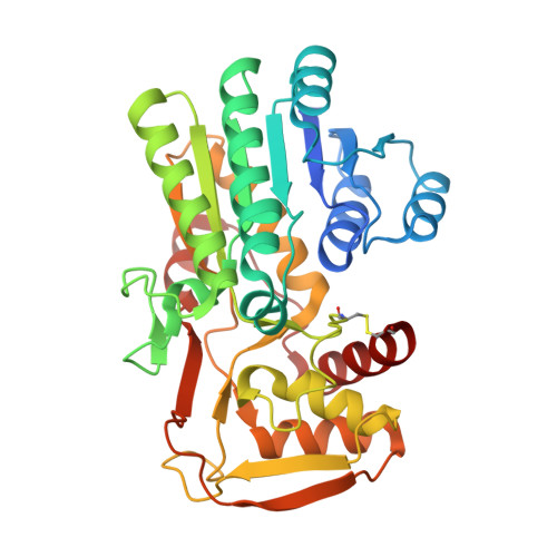 | |
UniProt | |||||
Entity Groups | |||||
| Sequence Clusters | 30% Identity50% Identity70% Identity90% Identity95% Identity100% Identity | ||||
| UniProt Group | Q0P9C3 | ||||
Sequence AnnotationsExpand | |||||
Reference Sequence | |||||
| Ligands 3 Unique | |||||
|---|---|---|---|---|---|
| ID | Chains | Name / Formula / InChI Key | 2D Diagram | 3D Interactions | |
| NAD (Subject of Investigation/LOI) Download:Ideal Coordinates CCD File | C [auth A], H [auth B] | NICOTINAMIDE-ADENINE-DINUCLEOTIDE C21 H27 N7 O14 P2 BAWFJGJZGIEFAR-NNYOXOHSSA-N |  | ||
| GOL Download:Ideal Coordinates CCD File | D [auth A] E [auth A] F [auth A] I [auth B] J [auth B] | GLYCEROL C3 H8 O3 PEDCQBHIVMGVHV-UHFFFAOYSA-N |  | ||
| ACT Download:Ideal Coordinates CCD File | G [auth A], M [auth B] | ACETATE ION C2 H3 O2 QTBSBXVTEAMEQO-UHFFFAOYSA-M |  | ||
| Length ( Å ) | Angle ( ˚ ) |
|---|---|
| a = 87.7 | α = 90 |
| b = 87.7 | β = 90 |
| c = 261.66 | γ = 90 |
| Software Name | Purpose |
|---|---|
| PHENIX | refinement |
| PDB_EXTRACT | data extraction |
| XDS | data reduction |
| SCALA | data scaling |
| PHASER | phasing |


