


Crystal structure of human insulin degrading enzyme (IDE) in complex with compound 3
Liang, W.G., Deprez, R., Bosc, D., Tang, W.To be published.
Experimental Data Snapshot
Starting Model: experimental
View more details
Entity ID: 1 | |||||
|---|---|---|---|---|---|
| Molecule | Chains | Sequence Length | Organism | Details | Image |
| Insulin-degrading enzyme | 990 | Homo sapiens | Mutation(s): 13 Gene Names: IDE EC: 3.4.24.56 | 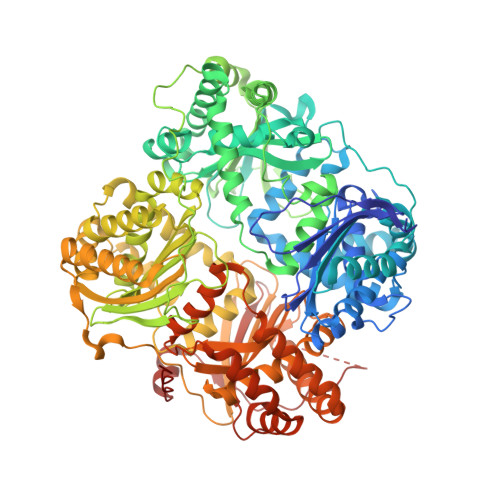 | |
UniProt & NIH Common Fund Data Resources | |||||
PHAROS: P14735 GTEx: ENSG00000119912 | |||||
Entity Groups | |||||
| Sequence Clusters | 30% Identity50% Identity70% Identity90% Identity95% Identity100% Identity | ||||
| UniProt Group | P14735 | ||||
Sequence AnnotationsExpand | |||||
Reference Sequence | |||||
| Ligands 4 Unique | |||||
|---|---|---|---|---|---|
| ID | Chains | Name / Formula / InChI Key | 2D Diagram | 3D Interactions | |
| VQG (Subject of Investigation/LOI) Download:Ideal Coordinates CCD File | D [auth A], J [auth B] | 3,4-difluoro-N-[(1S)-1-{1-[(2R)-4-(hydroxyamino)-4-oxo-1-(5,6,7,8-tetrahydronaphthalen-2-yl)butan-2-yl]-1H-1,2,3-triazol-4-yl}ethyl]benzamide C25 H27 F2 N5 O3 VQGQRXDQPMXXSZ-MGPUTAFESA-N |  | ||
| EPE Download:Ideal Coordinates CCD File | H [auth A], N [auth B] | 4-(2-HYDROXYETHYL)-1-PIPERAZINE ETHANESULFONIC ACID C8 H18 N2 O4 S JKMHFZQWWAIEOD-UHFFFAOYSA-N |  | ||
| DIO Download:Ideal Coordinates CCD File | E [auth A] F [auth A] G [auth A] K [auth B] L [auth B] | 1,4-DIETHYLENE DIOXIDE C4 H8 O2 RYHBNJHYFVUHQT-UHFFFAOYSA-N |  | ||
| ZN Download:Ideal Coordinates CCD File | C [auth A], I [auth B] | ZINC ION Zn PTFCDOFLOPIGGS-UHFFFAOYSA-N |  | ||
| Length ( Å ) | Angle ( ˚ ) |
|---|---|
| a = 267.88 | α = 90 |
| b = 267.88 | β = 90 |
| c = 89.611 | γ = 120 |
| Software Name | Purpose |
|---|---|
| PHENIX | refinement |
| HKL-3000 | data reduction |
| HKL-3000 | data scaling |
| PHASER | phasing |
| Funding Organization | Location | Grant Number |
|---|---|---|
| National Institutes of Health/National Institute of Mental Health (NIH/NIMH) | United States | R01 GM121964 |


