


Mapping the uncharted water channel of DHDPS
Board, A.J., Dobson, R.C.J.To be published.
Experimental Data Snapshot
Starting Model: experimental
View more details
Entity ID: 1 | |||||
|---|---|---|---|---|---|
| Molecule | Chains | Sequence Length | Organism | Details | Image |
| 4-hydroxy-tetrahydrodipicolinate synthase | 292 | Escherichia coli | Mutation(s): 1 Gene Names: dapA, A6V01_06140, A8C65_04230, ACU57_04510, AM464_03890, AML07_10805, APZ14_05880, AUQ13_12840, AUS26_08890, AW106_01920... EC: 4.3.3.7 | 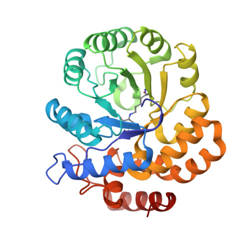 | |
UniProt | |||||
Entity Groups | |||||
| Sequence Clusters | 30% Identity50% Identity70% Identity90% Identity95% Identity100% Identity | ||||
| UniProt Group | P0A6L2 | ||||
Sequence AnnotationsExpand | |||||
Reference Sequence | |||||
| Ligands 3 Unique | |||||
|---|---|---|---|---|---|
| ID | Chains | Name / Formula / InChI Key | 2D Diagram | 3D Interactions | |
| LYS (Subject of Investigation/LOI) Download:Ideal Coordinates CCD File | D [auth A], H [auth B] | LYSINE C6 H15 N2 O2 KDXKERNSBIXSRK-YFKPBYRVSA-O |  | ||
| GOL Download:Ideal Coordinates CCD File | E [auth A], F [auth A], I [auth B] | GLYCEROL C3 H8 O3 PEDCQBHIVMGVHV-UHFFFAOYSA-N |  | ||
| K Download:Ideal Coordinates CCD File | C [auth A], G [auth B], J [auth B] | POTASSIUM ION K NPYPAHLBTDXSSS-UHFFFAOYSA-N |  | ||
| Modified Residues 1 Unique | |||||
|---|---|---|---|---|---|
| ID | Chains | Type | Formula | 2D Diagram | Parent |
| VPV Query on VPV | A, B | L-PEPTIDE LINKING | C13 H22 N2 O7 |  | LYS |
| Length ( Å ) | Angle ( ˚ ) |
|---|---|
| a = 121.299 | α = 90 |
| b = 121.299 | β = 90 |
| c = 109.781 | γ = 120 |
| Software Name | Purpose |
|---|---|
| REFMAC | refinement |
| Coot | model building |
| Aimless | data scaling |
| PHASER | phasing |


