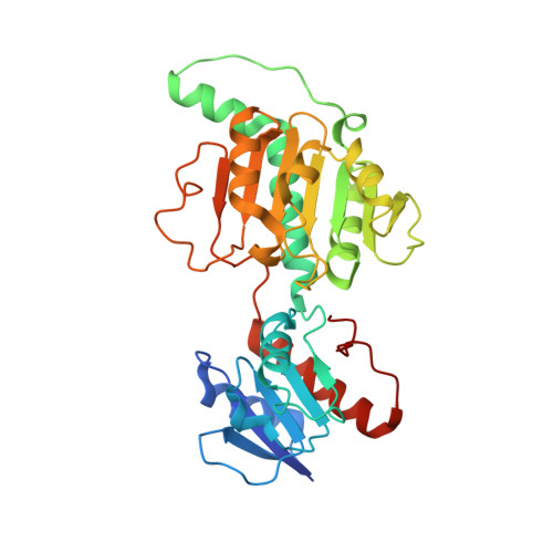
Structure of wild type Glyoxylate/Hydroxypyruvate reductase A from Escherichia Coli in complex with phosphate and NADP+
Vuksanovic, N., Silvaggi, N.R.To be published.
Experimental Data Snapshot
Starting Model: experimental
View more details
| Ligands 3 Unique | |||||
|---|---|---|---|---|---|
| ID | Chains | Name / Formula / InChI Key | 2D Diagram | 3D Interactions | |
| NAP Download:Ideal Coordinates CCD File | C [auth A] | NADP NICOTINAMIDE-ADENINE-DINUCLEOTIDE PHOSPHATE C21 H28 N7 O17 P3 XJLXINKUBYWONI-NNYOXOHSSA-N |  | ||
| PO4 Download:Ideal Coordinates CCD File | D [auth A], E [auth A], F [auth A] | PHOSPHATE ION O4 P NBIIXXVUZAFLBC-UHFFFAOYSA-K |  | ||
| GOL Download:Ideal Coordinates CCD File | B [auth A] | GLYCEROL C3 H8 O3 PEDCQBHIVMGVHV-UHFFFAOYSA-N |  | ||
| Length ( Å ) | Angle ( ˚ ) |
|---|---|
| a = 158.768 | α = 90 |
| b = 158.768 | β = 90 |
| c = 97.699 | γ = 120 |
| Software Name | Purpose |
|---|---|
| PHENIX | refinement |
| HKL-2000 | data reduction |
| HKL-2000 | data scaling |
| PHASER | phasing |