7JOM | pdb_00007jom

Crystal structure of Danio rerio histone deacetylase 6 catalytic domain 2 complexed with TO-317


Experimental Data Snapshot

  • Method: X-RAY DIFFRACTION
  • Resolution: 1.84 Å
  • R-Value Free: 
    0.207 (Depositor), 0.209 (DCC) 
  • R-Value Work: 
    0.168 (Depositor) 
  • R-Value Observed: 
    0.170 (Depositor) 

Starting Model: experimental
View more details

wwPDB Validation 3D Report Full Report

Validation slider image for 7JOM

Ligand Structure Quality Assessment 


This is version 1.2 of the entry. See complete history

Literature

Unique Molecular Interaction with the Histone Deacetylase 6 Catalytic Tunnel: Crystallographic and Biological Characterization of a Model Chemotype.

Olaoye, O.O.Watson, P.R.Nawar, N.Geletu, M.Sedighi, A.Bukhari, S.Raouf, Y.S.Manaswiyoungkul, P.Erdogan, F.Abdeldayem, A.Cabral, A.D.Hassan, M.M.Toutah, K.Shouksmith, A.E.Gawel, J.M.Israelian, J.Radu, T.B.Kachhiyapatel, N.de Araujo, E.D.Christianson, D.W.Gunning, P.T.

(2021) J Med Chem 64: 2691-2704

  • DOI: https://doi.org/10.1021/acs.jmedchem.0c01922
  • Primary Citation Related Structures: 
    7JOM

  • PubMed Abstract: 

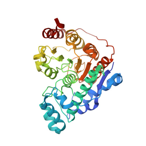
    Histone deacetylase 6 (HDAC6) is involved in multiple regulatory processes, ranging from cellular stress to intracellular transport. Inhibition of aberrant HDAC6 activity in several cancers and neurological diseases has been shown to be efficacious in both preclinical and clinical studies. While selective HDAC6 targeting has been pursued as an alternative to pan-HDAC drugs, identifying truly selective molecular templates has not been trivial. Herein, we report a structure-activity relationship study yielding TO-317 , which potently binds HDAC6 catalytic domain 2 ( K i = 0.7 nM) and inhibits the enzyme function (IC 50 = 2 nM). TO-317 exhibits 158-fold selectivity for HDAC6 over other HDAC isozymes by binding the catalytic Zn 2+ and, uniquely, making a never seen before direct hydrogen bond with the Zn 2+ coordinating residue, His614. This novel structural motif targeting the second-sphere His614 interaction, observed in a 1.84 Å resolution crystal structure with dr HDAC6 from zebrafish, can provide new pharmacophores for identifying enthalpically driven, high-affinity, HDAC6-selective inhibitors.


  • Organizational Affiliation
    • Department of Chemical and Physical Sciences, University of Toronto Mississauga, 3359 Mississauga Rd N., Mississauga, Ontario L5L 1C6, Canada.

Macromolecule Content 

  • Total Structure Weight: 82.21 kDa 
  • Atom Count: 5,962 
  • Modeled Residue Count: 709 
  • Deposited Residue Count: 728 
  • Unique protein chains: 1

Macromolecules

Find similar proteins by:|  3D Structure
Entity ID: 1
MoleculeChains  Sequence LengthOrganismDetailsImage
Hdac6 protein
A, B
364Danio rerioMutation(s): 0 
Gene Names: hdac6
EC: 3
UniProt
Find proteins for F8W4B7 (Danio rerio)
Explore F8W4B7 
Go to UniProtKB:  F8W4B7
Entity Groups
Sequence Clusters30% Identity50% Identity70% Identity90% Identity95% Identity100% Identity
UniProt GroupF8W4B7
Sequence Annotations
Expand
Reference Sequence

Small Molecules

Ligands 5 Unique
IDChains Name / Formula / InChI Key2D Diagram3D Interactions
TO3
(Subject of Investigation/LOI)

Query on TO3


Download:Ideal Coordinates CCD File
C [auth A],
M [auth B]
N-hydroxy-4-({[(pyridin-3-yl)methyl][(2,3,4,5-tetrafluorophenyl)sulfonyl]amino}methyl)benzamide
C20 H15 F4 N3 O4 S
HBOMISXRZUHCSH-UHFFFAOYSA-N
ZN

Query on ZN


Download:Ideal Coordinates CCD File
I [auth A],
P [auth B]
ZINC ION
Zn
PTFCDOFLOPIGGS-UHFFFAOYSA-N
EDO

Query on EDO


Download:Ideal Coordinates CCD File
D [auth A],
E [auth A],
N [auth B]
1,2-ETHANEDIOL
C2 H6 O2
LYCAIKOWRPUZTN-UHFFFAOYSA-N
FMT

Query on FMT


Download:Ideal Coordinates CCD File
F [auth A],
G [auth A],
H [auth A],
O [auth B]
FORMIC ACID
C H2 O2
BDAGIHXWWSANSR-UHFFFAOYSA-N
K

Query on K


Download:Ideal Coordinates CCD File
J [auth A],
K [auth A],
L [auth A],
Q [auth B],
R [auth B]
POTASSIUM ION
K
NPYPAHLBTDXSSS-UHFFFAOYSA-N

Experimental Data & Validation

Experimental Data

  • Method: X-RAY DIFFRACTION
  • Resolution: 1.84 Å
  • R-Value Free:  0.207 (Depositor), 0.209 (DCC) 
  • R-Value Work:  0.168 (Depositor) 
  • R-Value Observed: 0.170 (Depositor) 
Space Group: P 21 21 21
Unit Cell:
Length ( Å )Angle ( ˚ )
a = 75.46α = 90
b = 96.45β = 90
c = 96.45γ = 90
Software Package:
Software NamePurpose
PHENIXrefinement
iMOSFLMdata reduction
Aimlessdata scaling
PHASERphasing

Structure Validation

View Full Validation Report


Ligand Structure Quality Assessment 


Entry History 

& Funding Information

Deposition Data


Funding OrganizationLocationGrant Number
National Institutes of Health/National Institute of General Medical Sciences (NIH/NIGMS)United StatesGM49758

Revision History  (Full details and data files)

  • Version 1.0: 2021-06-16
    Type: Initial release
  • Version 1.1: 2021-09-01
    Changes: Database references
  • Version 1.2: 2023-10-18
    Changes: Data collection, Refinement description