7JMD

Sheep Connexin-46 at 2.5 angstroms resolution, Lipid Class 1


Experimental Data Snapshot

  • Method: ELECTRON MICROSCOPY
  • Resolution: 2.50 Å
  • Aggregation State: PARTICLE 
  • Reconstruction Method: SINGLE PARTICLE 

wwPDB Validation   3D Report Full Report


This is version 1.0 of the entry. See complete history


Literature

Connexin-46/50 in a dynamic lipid environment resolved by CryoEM at 1.9 angstrom.

Flores, J.A.Haddad, B.G.Dolan, K.A.Myers, J.B.Yoshioka, C.C.Copperman, J.Zuckerman, D.M.Reichow, S.L.

(2020) Nat Commun 11: 4331-4331

  • DOI: https://doi.org/10.1038/s41467-020-18120-5
  • Primary Citation of Related Structures:  
    7JJP, 7JKC, 7JLW, 7JM9, 7JMC, 7JMD, 7JN0, 7JN1

  • PubMed Abstract: 

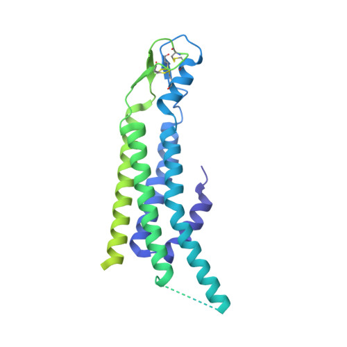
    Gap junctions establish direct pathways for cells to transfer metabolic and electrical messages. The local lipid environment is known to affect the structure, stability and intercellular channel activity of gap junctions; however, the molecular basis for these effects remains unknown. Here, we incorporate native connexin-46/50 (Cx46/50) intercellular channels into a dual lipid nanodisc system, mimicking a native cell-to-cell junction. Structural characterization by CryoEM reveals a lipid-induced stabilization to the channel, resulting in a 3D reconstruction at 1.9 Å resolution. Together with all-atom molecular dynamics simulations, it is shown that Cx46/50 in turn imparts long-range stabilization to the dynamic local lipid environment that is specific to the extracellular lipid leaflet. In addition, ~400 water molecules are resolved in the CryoEM map, localized throughout the intercellular permeation pathway and contributing to the channel architecture. These results illustrate how the aqueous-lipid environment is integrated with the architectural stability, structure and function of gap junction communication channels.


  • Organizational Affiliation

    Department of Chemical Physiology and Biochemistry, Oregon Health and Science University, Portland, OR, 97239, USA.


Macromolecules
Find similar proteins by:  (by identity cutoff)  |  3D Structure
Entity ID: 1
MoleculeChains Sequence LengthOrganismDetailsImage
Gap junction alpha-3 protein
A, B, C, D, E
A, B, C, D, E, F, G, H, I, J, K, L
413Ovis ariesMutation(s): 0 
Membrane Entity: Yes 
UniProt
Find proteins for Q9TU17 (Ovis aries)
Explore Q9TU17 
Go to UniProtKB:  Q9TU17
Entity Groups  
Sequence Clusters30% Identity50% Identity70% Identity90% Identity95% Identity100% Identity
UniProt GroupQ9TU17
Sequence Annotations
Expand
  • Reference Sequence
Small Molecules
Ligands 1 Unique
IDChains Name / Formula / InChI Key2D Diagram3D Interactions
MC3 (Subject of Investigation/LOI)
Query on MC3

Download Ideal Coordinates CCD File 
AA [auth B]
AB [auth D]
AC [auth G]
AD [auth I]
AE [auth K]
AA [auth B],
AB [auth D],
AC [auth G],
AD [auth I],
AE [auth K],
BA [auth B],
BB [auth D],
BC [auth G],
BD [auth I],
BE [auth K],
CA [auth B],
CB [auth D],
CC [auth G],
CD [auth I],
CE [auth K],
DA [auth B],
DB [auth D],
DC [auth G],
DD [auth I],
DE [auth L],
EA [auth B],
EB [auth E],
EC [auth G],
ED [auth I],
EE [auth L],
FA [auth B],
FB [auth E],
FC [auth G],
FD [auth I],
FE [auth L],
GA [auth B],
GB [auth E],
GC [auth G],
GD [auth I],
GE [auth L],
HA [auth B],
HB [auth E],
HC [auth G],
HD [auth J],
HE [auth L],
IA [auth C],
IB [auth E],
IC [auth G],
ID [auth J],
IE [auth L],
JA [auth C],
JB [auth E],
JC [auth G],
JD [auth J],
JE [auth L],
KA [auth C],
KB [auth E],
KC [auth G],
KD [auth J],
KE [auth L],
LA [auth C],
LB [auth E],
LC [auth H],
LD [auth J],
LE [auth L],
M [auth A],
MA [auth C],
MB [auth E],
MC [auth H],
MD [auth J],
ME [auth L],
N [auth A],
NA [auth C],
NB [auth E],
NC [auth H],
ND [auth J],
NE [auth L],
O [auth A],
OA [auth C],
OB [auth E],
OC [auth H],
OD [auth J],
P [auth A],
PA [auth C],
PB [auth F],
PC [auth H],
PD [auth J],
Q [auth A],
QA [auth C],
QB [auth F],
QC [auth H],
QD [auth J],
R [auth A],
RA [auth C],
RB [auth F],
RC [auth H],
RD [auth J],
S [auth A],
SA [auth C],
SB [auth F],
SC [auth H],
SD [auth K],
T [auth A],
TA [auth D],
TB [auth F],
TC [auth H],
TD [auth K],
U [auth A],
UA [auth D],
UB [auth F],
UC [auth H],
UD [auth K],
V [auth A],
VA [auth D],
VB [auth F],
VC [auth H],
VD [auth K],
W [auth A],
WA [auth D],
WB [auth F],
WC [auth I],
WD [auth K],
X [auth B],
XA [auth D],
XB [auth F],
XC [auth I],
XD [auth K],
Y [auth B],
YA [auth D],
YB [auth F],
YC [auth I],
YD [auth K],
Z [auth B],
ZA [auth D],
ZB [auth F],
ZC [auth I],
ZD [auth K]
1,2-DIMYRISTOYL-RAC-GLYCERO-3-PHOSPHOCHOLINE
C36 H72 N O8 P
CITHEXJVPOWHKC-UUWRZZSWSA-N
Experimental Data & Validation

Experimental Data

  • Method: ELECTRON MICROSCOPY
  • Resolution: 2.50 Å
  • Aggregation State: PARTICLE 
  • Reconstruction Method: SINGLE PARTICLE 
EM Software:
TaskSoftware PackageVersion
RECONSTRUCTIONRELION3.0
MODEL REFINEMENTPHENIX

Structure Validation

View Full Validation Report


Entry History & Funding Information

Deposition Data


Funding OrganizationLocationGrant Number
National Institutes of Health/National Eye Institute (NIH/NEI)United StatesR35GM124779
National Institutes of Health/National Eye Institute (NIH/NEI)United States1F31EY030409-01

Revision History  (Full details and data files)

  • Version 1.0: 2020-09-09
    Type: Initial release