7JIU

HUMAN PI3KDELTA IN COMPLEX WITH COMPOUND 2F


Experimental Data Snapshot

  • Method: X-RAY DIFFRACTION
  • Resolution: 2.12 Å
  • R-Value Free: 0.246 
  • R-Value Work: 0.212 
  • R-Value Observed: 0.213 

wwPDB Validation   3D Report Full Report


Ligand Structure Quality Assessment 


This is version 1.2 of the entry. See complete history


Literature

Optimization of Versatile Oxindoles as Selective PI3K delta Inhibitors.

Methot, J.L.Achab, A.Christopher, M.Zhou, H.McGowan, M.A.Trotter, B.W.Fradera, X.Lesburg, C.A.Goldenblatt, P.Hill, A.Chen, D.Otte, K.M.Augustin, M.Shah, S.Katz, J.D.

(2020) ACS Med Chem Lett 11: 2461-2469

  • DOI: https://doi.org/10.1021/acsmedchemlett.0c00441
  • Primary Citation of Related Structures:  
    7JIS, 7JIU

  • PubMed Abstract: 

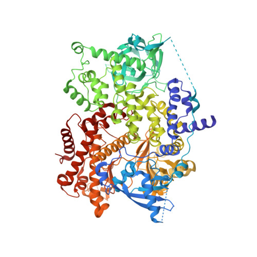
    The 3,3-disubstituted oxindole moiety is a versatile and rigid three-dimensionally shaped scaffold. When engineered with a purine hinge-binding core, exceptionally selective PI3Kδ kinase inhibitors were discovered by exploiting small differences in isoform selectivity pockets. Crystal structures of early lead 2f bound to PI3Kδ and PI3Kα helped rationalize the high selectivity observed with 2f . By attenuating the lypophilicity and metabolic liabilities of an oxindole moiety, we improved the preclinical species PK and solubility and reduced adenosine uptake activity. The excellent potency and kinome selectivity of 7-azaoxindole 4d and spirooxindole 5d , together with a low plasma clearance and good half-life in rat and dog, supported a low once-daily predicted human dose.


  • Organizational Affiliation

    Discovery Chemistry, Computational and Structural Chemistry, In Vitro Pharmacology, Pharmacokinetics, Pharmacodynamics and Drug Metabolism, Merck & Co., Inc., Boston, Massachusetts 02115, United States.


Macromolecules
Find similar proteins by:  (by identity cutoff)  |  3D Structure
Entity ID: 1
MoleculeChains Sequence LengthOrganismDetailsImage
Phosphatidylinositol 4,5-bisphosphate 3-kinase catalytic subunit alpha isoform946Homo sapiensMutation(s): 0 
EC: 2.7.1.153 (PDB Primary Data), 2.7.11.1 (PDB Primary Data)
UniProt & NIH Common Fund Data Resources
Find proteins for P42336 (Homo sapiens)
Explore P42336 
Go to UniProtKB:  P42336
PHAROS:  P42336
GTEx:  ENSG00000121879 
Entity Groups  
Sequence Clusters30% Identity50% Identity70% Identity90% Identity95% Identity100% Identity
UniProt GroupP42336
Sequence Annotations
Expand
  • Reference Sequence
Small Molecules
Ligands 1 Unique
IDChains Name / Formula / InChI Key2D Diagram3D Interactions
VBS (Subject of Investigation/LOI)
Query on VBS

Download Ideal Coordinates CCD File 
B [auth A](3S)-3-benzyl-5-[9-ethyl-8-(2-methylpyrimidin-5-yl)-9H-purin-6-yl]-3-methyl-1,3-dihydro-2H-indol-2-one
C28 H25 N7 O
FFRRAJYYORCXFZ-NDEPHWFRSA-N
Experimental Data & Validation

Experimental Data

  • Method: X-RAY DIFFRACTION
  • Resolution: 2.12 Å
  • R-Value Free: 0.246 
  • R-Value Work: 0.212 
  • R-Value Observed: 0.213 
  • Space Group: P 21 21 21
Unit Cell:
Length ( Å )Angle ( ˚ )
a = 58.457α = 90
b = 135.132β = 90
c = 142.045γ = 90
Software Package:
Software NamePurpose
XSCALEdata scaling
REFMACrefinement
PDB_EXTRACTdata extraction
XDSdata reduction
MOLREPphasing

Structure Validation

View Full Validation Report


Ligand Structure Quality Assessment 


Entry History 

Deposition Data

Revision History  (Full details and data files)

  • Version 1.0: 2021-06-30
    Type: Initial release
  • Version 1.1: 2024-03-06
    Changes: Advisory, Data collection, Database references
  • Version 1.2: 2024-04-03
    Changes: Refinement description