7JID | pdb_00007jid

Crystal structure of the L780 UDP-rhamnose synthase from Acanthamoeba polyphaga mimivirus


Experimental Data Snapshot

  • Method: X-RAY DIFFRACTION
  • Resolution: 1.45 Å
  • R-Value Free: 
    0.227 (Depositor), 0.233 (DCC) 
  • R-Value Work: 
    0.198 (Depositor), 0.206 (DCC) 
  • R-Value Observed: 
    0.199 (Depositor) 

Starting Model: experimental
View more details

wwPDB Validation 3D Report Full Report

Validation slider image for 7JID

Ligand Structure Quality Assessment 


This is version 1.2 of the entry. See complete history

Literature

The high-resolution structure of a UDP-L-rhamnose synthase from Acanthamoeba polyphaga Mimivirus.

Bockhaus, N.J.Ferek, J.D.Thoden, J.B.Holden, H.M.

(2020) Protein Sci 29: 2164-2174

  • DOI: https://doi.org/10.1002/pro.3928
  • Primary Citation Related Structures: 
    7JID

  • PubMed Abstract: 

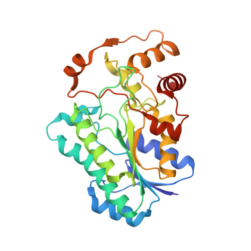
    For the field of virology, perhaps one of the most paradigm-shifting events so far in the 21st century was the identification of the giant double-stranded DNA virus that infects amoebae. Remarkably, this virus, known as Mimivirus, has a genome that encodes for nearly 1,000 proteins, some of which are involved in the biosynthesis of unusual sugars. Indeed, the virus is coated by a layer of glycosylated fibers that contain d-glucose, N-acetyl-d-glucosamine, l-rhamnose, and 4-amino-4,6-dideoxy-d-glucose. Here we describe a combined structural and enzymological investigation of the protein encoded by the open-reading frame L780, which corresponds to an l-rhamnose synthase. The structure of the L780/NADP + /UDP-l-rhamnose ternary complex was determined to 1.45 Å resolution and refined to an overall R-factor of 19.9%. Each subunit of the dimeric protein adopts a bilobal-shaped appearance with the N-terminal domain harboring the dinucleotide-binding site and the C-terminal domain positioning the UDP-sugar into the active site. The overall molecular architecture of L780 places it into the short-chain dehydrogenase/reductase superfamily. Kinetic analyses indicate that the enzyme can function on either UDP- and dTDP-sugars but displays a higher catalytic efficiency with the UDP-linked substrate. Site-directed mutagenesis experiments suggest that both Cys 108 and Lys 175 play key roles in catalysis. This structure represents the first model of a viral UDP-l-rhamnose synthase and provides new details into these fascinating enzymes.


  • Organizational Affiliation
    • Department of Biochemistry, University of Wisconsin, Madison, Wisconsin, USA.

Macromolecule Content 

  • Total Structure Weight: 69.53 kDa 
  • Atom Count: 5,532 
  • Modeled Residue Count: 574 
  • Deposited Residue Count: 584 
  • Unique protein chains: 1

Macromolecules

Find similar proteins by:|  3D Structure
Entity ID: 1
MoleculeChains  Sequence LengthOrganismDetailsImage
UDP-L-Rhamnose Synthase
A, B
292Acanthamoeba polyphaga mimivirusMutation(s): 0 
Gene Names: MIMI_L780
UniProt
Find proteins for Q5UPS5 (Acanthamoeba polyphaga mimivirus)
Explore Q5UPS5 
Go to UniProtKB:  Q5UPS5
Entity Groups
Sequence Clusters30% Identity50% Identity70% Identity90% Identity95% Identity100% Identity
UniProt GroupQ5UPS5
Sequence Annotations
Expand
Reference Sequence

Small Molecules

Ligands 3 Unique
IDChains Name / Formula / InChI Key2D Diagram3D Interactions
NAP

Query on NAP


Download:Ideal Coordinates CCD File
D [auth A],
L [auth B]
NADP NICOTINAMIDE-ADENINE-DINUCLEOTIDE PHOSPHATE
C21 H28 N7 O17 P3
XJLXINKUBYWONI-NNYOXOHSSA-N
AWU
(Subject of Investigation/LOI)

Query on AWU


Download:Ideal Coordinates CCD File
C [auth A],
K [auth B]
[[(2~{R},3~{S},4~{R},5~{R})-5-[2,4-bis(oxidanylidene)pyrimidin-1-yl]-3,4-bis(oxidanyl)oxolan-2-yl]methoxy-oxidanyl-phosphoryl] [(2~{R},3~{R},4~{R},5~{R},6~{S})-6-methyl-3,4,5-tris(oxidanyl)oxan-2-yl] hydrogen phosphate
C15 H24 N2 O16 P2
DRDCJEIZVLVWNC-SLBWPEPYSA-N
EDO

Query on EDO


Download:Ideal Coordinates CCD File
E [auth A]
F [auth A]
G [auth A]
H [auth A]
I [auth A]
E [auth A],
F [auth A],
G [auth A],
H [auth A],
I [auth A],
J [auth A],
M [auth B],
N [auth B],
O [auth B]
1,2-ETHANEDIOL
C2 H6 O2
LYCAIKOWRPUZTN-UHFFFAOYSA-N

Experimental Data & Validation

Experimental Data

  • Method: X-RAY DIFFRACTION
  • Resolution: 1.45 Å
  • R-Value Free:  0.227 (Depositor), 0.233 (DCC) 
  • R-Value Work:  0.198 (Depositor), 0.206 (DCC) 
  • R-Value Observed: 0.199 (Depositor) 
Space Group: P 1
Unit Cell:
Length ( Å )Angle ( ˚ )
a = 49.803α = 81.94
b = 52.989β = 88.68
c = 70.149γ = 66.74
Software Package:
Software NamePurpose
REFMACrefinement
PDB_EXTRACTdata extraction
SAINTdata reduction
SADABSdata scaling
PHASERphasing

Structure Validation

View Full Validation Report


Ligand Structure Quality Assessment 


Entry History 

& Funding Information

Deposition Data


Funding OrganizationLocationGrant Number
National Institutes of Health/National Institute of General Medical Sciences (NIH/NIGMS)United StatesGM134643

Revision History  (Full details and data files)

  • Version 1.0: 2020-08-26
    Type: Initial release
  • Version 1.1: 2020-11-04
    Changes: Database references
  • Version 1.2: 2023-10-18
    Changes: Data collection, Database references, Refinement description