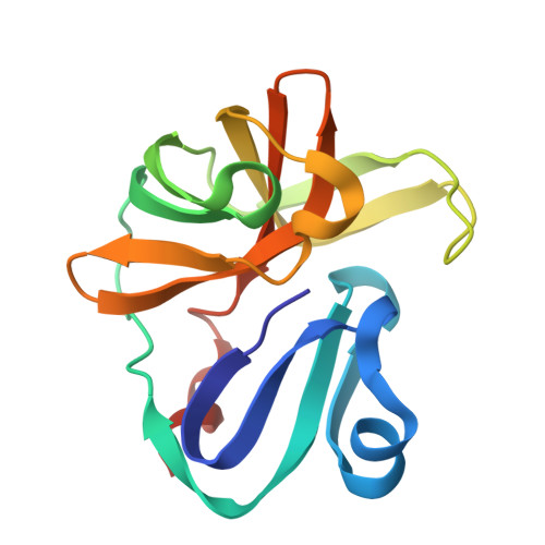
Group deposition of Coxsackievirus A16 (G-10) 2A protease in complex with inhibitors from the ASAP AViDD centre
Lithgo, R.M., Fairhead, M., Koekemoer, L., Balcomb, B.H., Capkin, E., Chandran, A.V., Golding, M., Godoy, A.S., Aschenbrenner, J.C., Marples, P.G., Ni, X., Thompson, W., Tomlinson, C.W.E., Wild, C., Winokan, M., Xavier, M.-A.E., Kenton, N., Tucker, J., DiPoto, M., Lee, A., Fearon, D., von Delft, F.To be published.