7GSL | pdb_00007gsl

PanDDA Analysis group deposition -- Crystal structure of PTP1B in complex with FMSOA000274b

  • Classification: HYDROLASE
  • Organism(s): Homo sapiens
  • Expression System: Escherichia coli
  • Mutation(s): Yes 

  • Deposited: 2024-01-03 Released: 2024-01-24 
  • Deposition Author(s): Mehlman, T., Ginn, H.M., Keedy, D.A.
  • Funding Organization(s): National Institutes of Health/National Institute of General Medical Sciences (NIH/NIGMS), Helmholtz Association

Experimental Data Snapshot

  • Method: X-RAY DIFFRACTION
  • Resolution: 1.77 Å
  • R-Value Free: 
    0.226 (Depositor), 0.228 (DCC) 
  • R-Value Work: 
    0.191 (Depositor) 
  • R-Value Observed: 
    0.192 (Depositor) 

wwPDB Validation 3D Report Full Report

Validation slider image for 7GSL

Ligand Structure Quality Assessment 


This is version 1.2 of the entry. See complete history

Literature

An expanded trove of fragment-bound structures for the allosteric enzyme PTP1B from computational reanalysis of large-scale crystallographic data.

Mehlman, T.Ginn, H.M.Keedy, D.A.

(2024) Structure 32: 1231-1238.e4

  • DOI: https://doi.org/10.1016/j.str.2024.05.010
  • Primary Citation Related Structures: 
    7GS7, 7GS8, 7GS9, 7GSA, 7GSB, 7GSC, 7GSD, 7GSE, 7GSF, 7GSG, 7GSH, 7GSI, 7GSJ, 7GSK, 7GSL, 7GSM, 7GSN, 7GSO, 7GSQ, 7GSR, 7GST, 7GSU, 7GSV, 7GSW, 7GSX, 7GSY, 7GSZ, 7GT0, 7GT1, 7GT2, 7GT3, 7GT4, 7GT5, 7GT6, 7GT7, 7GT8, 7GT9, 7GTA, 7GTB, 7GTC, 7GTD, 7GTE, 7GTF, 7GTG, 7GTH, 7GTI, 7GTJ, 7GTK, 7GTL, 7GTM, ... Search all related entries

  • PubMed Abstract: 

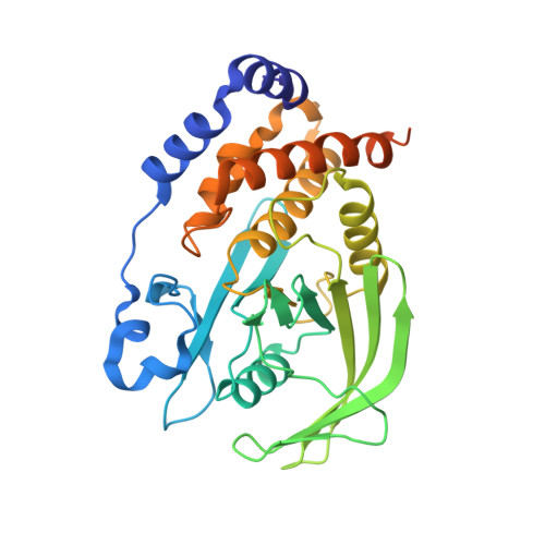
    Due to their low binding affinities, detecting small-molecule fragments bound to protein structures from crystallographic datasets has been a challenge. Here, we report a trove of 65 new fragment hits for PTP1B, an "undruggable" therapeutic target enzyme for diabetes and cancer. These structures were obtained from computational analysis of data from a large crystallographic screen, demonstrating the power of this approach to elucidate many (∼50% more) "hidden" ligand-bound states of proteins. Our new structures include a fragment hit found in a novel binding site in PTP1B with a unique location relative to the active site, one that links adjacent allosteric sites, and, perhaps most strikingly, a fragment that induces long-range allosteric protein conformational responses. Altogether, our research highlights the utility of computational analysis of crystallographic data, makes publicly available dozens of new ligand-bound structures of a high-value drug target, and identifies novel aspects of ligandability and allostery in PTP1B.


  • Organizational Affiliation
    • Structural Biology Initiative, CUNY Advanced Science Research Center, New York, NY 10031, USA; PhD Program in Biochemistry, CUNY Graduate Center, New York, NY 10016, USA.

Macromolecule Content 

  • Total Structure Weight: 37.64 kDa 
  • Atom Count: 2,582 
  • Modeled Residue Count: 284 
  • Deposited Residue Count: 321 
  • Unique protein chains: 1

Macromolecules

Find similar proteins by:|  3D Structure
Entity ID: 1
MoleculeChains  Sequence LengthOrganismDetailsImage
Tyrosine-protein phosphatase non-receptor type 1321Homo sapiensMutation(s): 2 
Gene Names: PTPN1PTP1B
EC: 3.1.3.48
UniProt & NIH Common Fund Data Resources
Find proteins for P18031 (Homo sapiens)
Explore P18031 
Go to UniProtKB:  P18031
PHAROS:  P18031
GTEx:  ENSG00000196396 
Entity Groups
Sequence Clusters30% Identity50% Identity70% Identity90% Identity95% Identity100% Identity
UniProt GroupP18031
Sequence Annotations
Expand
Reference Sequence

Small Molecules

Ligands 2 Unique
IDChains Name / Formula / InChI Key2D Diagram3D Interactions
A1AA8
(Subject of Investigation/LOI)

Query on A1AA8


Download:Ideal Coordinates CCD File
C [auth A]2-(methylsulfanyl)pyridine-3-carboxamide
C7 H8 N2 O S
YDYXNHSKBIZKFD-UHFFFAOYSA-N
TRS

Query on TRS


Download:Ideal Coordinates CCD File
B [auth A]2-AMINO-2-HYDROXYMETHYL-PROPANE-1,3-DIOL
C4 H12 N O3
LENZDBCJOHFCAS-UHFFFAOYSA-O

Experimental Data & Validation

Experimental Data

  • Method: X-RAY DIFFRACTION
  • Resolution: 1.77 Å
  • R-Value Free:  0.226 (Depositor), 0.228 (DCC) 
  • R-Value Work:  0.191 (Depositor) 
  • R-Value Observed: 0.192 (Depositor) 
Space Group: P 31 2 1
Unit Cell:
Length ( Å )Angle ( ˚ )
a = 89.944α = 90
b = 89.944β = 90
c = 106.588γ = 120
Software Package:
Software NamePurpose
REFMACrefinement
XSCALEdata scaling
PDB_EXTRACTdata extraction
PHASERphasing
XDSdata reduction

Structure Validation

View Full Validation Report


Ligand Structure Quality Assessment 


Entry History 

& Funding Information

Deposition Data


Funding OrganizationLocationGrant Number
National Institutes of Health/National Institute of General Medical Sciences (NIH/NIGMS)United StatesGM133769
Helmholtz AssociationGermanyVH-NG-19-02

Revision History  (Full details and data files)

  • Version 1.0: 2024-01-24
    Type: Initial release
  • Version 1.1: 2024-04-24
    Changes: Database references
  • Version 1.2: 2025-12-10
    Changes: Database references, Structure summary