


PanDDA analysis group deposition
Kumar, A., Knapp, S., Structural Genomics Consortium (SGC)To be published.
Experimental Data Snapshot
Starting Model: experimental
View more details
Entity ID: 1 | |||||
|---|---|---|---|---|---|
| Molecule | Chains | Sequence Length | Organism | Details | Image |
| Microtubule-associated proteins 1A/1B light chain 3B | 121 | Homo sapiens | Mutation(s): 0 Gene Names: MAP1LC3B, MAP1ALC3 | 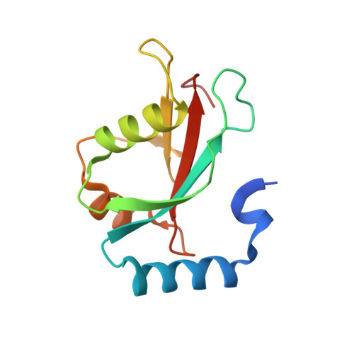 | |
UniProt & NIH Common Fund Data Resources | |||||
PHAROS: Q9GZQ8 GTEx: ENSG00000140941 | |||||
Entity Groups | |||||
| Sequence Clusters | 30% Identity50% Identity70% Identity90% Identity95% Identity100% Identity | ||||
| UniProt Group | Q9GZQ8 | ||||
Sequence AnnotationsExpand | |||||
Reference Sequence | |||||
| Ligands 3 Unique | |||||
|---|---|---|---|---|---|
| ID | Chains | Name / Formula / InChI Key | 2D Diagram | 3D Interactions | |
| K0J (Subject of Investigation/LOI) Download:Ideal Coordinates CCD File | B [auth A] | N-ethyl-1H-1,2,3-triazole-4-carboxamide C5 H8 N4 O VTJBQJOBKLNCDW-UHFFFAOYSA-N |  | ||
| EDO Download:Ideal Coordinates CCD File | C [auth A] D [auth A] E [auth A] F [auth A] G [auth A] | 1,2-ETHANEDIOL C2 H6 O2 LYCAIKOWRPUZTN-UHFFFAOYSA-N |  | ||
| CL Download:Ideal Coordinates CCD File | L [auth A] | CHLORIDE ION Cl VEXZGXHMUGYJMC-UHFFFAOYSA-M |  | ||
| Length ( Å ) | Angle ( ˚ ) |
|---|---|
| a = 61.18 | α = 90 |
| b = 61.18 | β = 90 |
| c = 35.47 | γ = 90 |
| Software Name | Purpose |
|---|---|
| REFMAC | refinement |
| Aimless | data scaling |
| PDB_EXTRACT | data extraction |
| XDS | data reduction |
| REFMAC | phasing |
| Funding Organization | Location | Grant Number |
|---|---|---|
| European Union (EU) | European Union | 875510 |


