


Crystal Structure of a rat Autotaxin complex
Hunziker, D., Joachim, S.C., Ullmer, C., Rudolph, M.G.To be published.
Experimental Data Snapshot
Starting Model: experimental
View more details
Entity ID: 1 | |||||
|---|---|---|---|---|---|
| Molecule | Chains | Sequence Length | Organism | Details | Image |
| Isoform 2 of Ectonucleotide pyrophosphatase/phosphodiesterase family member 2 | 846 | Rattus norvegicus | Mutation(s): 3 Gene Names: Enpp2, Atx, Npps2 EC: 3.1.4.39 (PDB Primary Data), 3.1.4.4 (UniProt) | 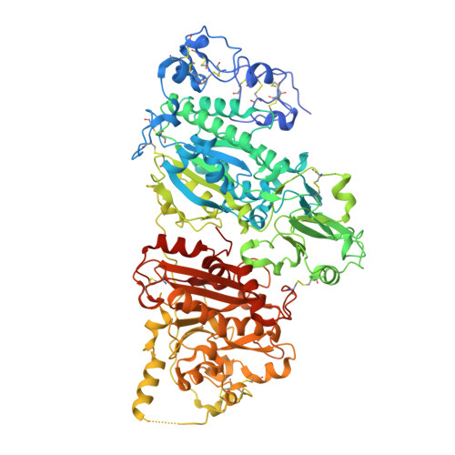 | |
UniProt | |||||
Entity Groups | |||||
| Sequence Clusters | 30% Identity50% Identity70% Identity90% Identity95% Identity100% Identity | ||||
| UniProt Group | Q64610 | ||||
Glycosylation | |||||
| Glycosylation Sites: 1 | Go to GlyGen: Q64610-1 | ||||
Sequence AnnotationsExpand | |||||
Reference Sequence | |||||
Entity ID: 2 | |||||
|---|---|---|---|---|---|
| Molecule | Chains | Length | 2D Diagram | Glycosylation | D Interactions |
| alpha-D-mannopyranose-(1-2)-alpha-D-mannopyranose-(1-3)-alpha-D-mannopyranose-(1-6)-[alpha-D-mannopyranose-(1-2)-alpha-D-mannopyranose-(1-3)]beta-D-mannopyranose-(1-4)-2-acetamido-2-deoxy-beta-D-glucopyranose-(1-4)-2-acetamido-2-deoxy-beta-D-glucopyranose | B | 8 |  | N-Glycosylation | |
Glycosylation Resources | |||||
GlyTouCan: G33303XH GlyCosmos: G33303XH GlyGen: G33303XH | |||||
| Ligands 7 Unique | |||||
|---|---|---|---|---|---|
| ID | Chains | Name / Formula / InChI Key | 2D Diagram | 3D Interactions | |
| XW9 (Subject of Investigation/LOI) Download:Ideal Coordinates CCD File | C [auth A] | [5-{2-cyclopropyl-6-[(oxan-4-yl)methoxy]pyridine-4-carbonyl}-3,4,5,6-tetrahydropyrrolo[3,4-c]pyrrol-2(1H)-yl](3-hydroxy-4,7-dihydro[1,2]oxazolo[5,4-c]pyridin-6(5H)-yl)methanone C28 H33 N5 O6 IFTRMTXDKJQHOD-UHFFFAOYSA-N |  | ||
| ZN Download:Ideal Coordinates CCD File | G [auth A] | ZINC ION Zn PTFCDOFLOPIGGS-UHFFFAOYSA-N |  | ||
| ACT Download:Ideal Coordinates CCD File | E [auth A] | ACETATE ION C2 H3 O2 QTBSBXVTEAMEQO-UHFFFAOYSA-M |  | ||
| CA Download:Ideal Coordinates CCD File | I [auth A] | CALCIUM ION Ca BHPQYMZQTOCNFJ-UHFFFAOYSA-N |  | ||
| K Download:Ideal Coordinates CCD File | D [auth A] | POTASSIUM ION K NPYPAHLBTDXSSS-UHFFFAOYSA-N |  | ||
| CL Download:Ideal Coordinates CCD File | F [auth A] | CHLORIDE ION Cl VEXZGXHMUGYJMC-UHFFFAOYSA-M |  | ||
| NA Download:Ideal Coordinates CCD File | H [auth A] | SODIUM ION Na FKNQFGJONOIPTF-UHFFFAOYSA-N |  | ||
| Length ( Å ) | Angle ( ˚ ) |
|---|---|
| a = 84.125 | α = 90 |
| b = 91.651 | β = 90 |
| c = 120.236 | γ = 90 |
| Software Name | Purpose |
|---|---|
| XSCALE | data scaling |
| REFMAC | refinement |
| PDB_EXTRACT | data extraction |
| XDS | data reduction |
| PHASER | phasing |
| Funding Organization | Location | Grant Number |
|---|---|---|
| F. Hoffmann-La Roche LTD | Switzerland | -- |


