7FVS | pdb_00007fvs

Crystal Structure of S. aureus gyrase in complex with 4-[[1-[(1-chloro-6,7-dihydro-5H-cyclopenta[c]pyridin-6-yl)methyl]azetidin-3-yl]methylamino]-6-fluorochromen-2-one


Experimental Data Snapshot

  • Method: X-RAY DIFFRACTION
  • Resolution: 2.16 Å
  • R-Value Free: 
    0.221 (Depositor), 0.200 (DCC) 
  • R-Value Work: 
    0.185 (Depositor), 0.175 (DCC) 
  • R-Value Observed: 
    0.191 (Depositor) 

Starting Model: experimental
View more details

wwPDB Validation 3D Report Full Report

Validation slider image for 7FVS

Ligand Structure Quality Assessment 


This is version 1.1 of the entry. See complete history

Literature

Discovery of a Series of Indane-Containing NBTIs with Activity against Multidrug-Resistant Gram-Negative Pathogens.

Cumming, J.G.Kreis, L.Kuhne, H.Wermuth, R.Vercruysse, M.Kramer, C.Rudolph, M.G.Xu, Z.

(2023) ACS Med Chem Lett 14: 993-998

  • DOI: https://doi.org/10.1021/acsmedchemlett.3c00187
  • Primary Citation Related Structures: 
    7FVS, 7FVT

  • PubMed Abstract: 

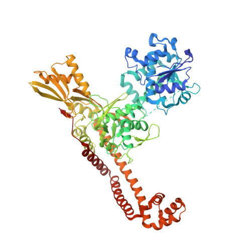
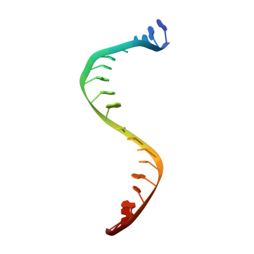
    The rise of multidrug-resistant (MDR) Gram-negative bacteria is a major global health problem necessitating the discovery of new classes of antibiotics. Novel bacterial topoisomerase inhibitors (NBTIs) target the clinically validated bacterial type II topoisomerases with a distinct binding site and mechanism of action to fluoroquinolone antibiotics, thus avoiding cross-resistance to this drug class. Here we report the discovery of a series of NBTIs incorporating a novel indane DNA binding moiety. X-ray cocrystal structures of compounds 2 and 17a bound to Staphylococcus aureus DNA gyrase-DNA were determined, revealing specific interactions with the enzyme binding pocket at the GyrA dimer interface and a long-range electrostatic interaction between the basic amine in the linker and the carboxylate of Asp83. Exploration of the structure-activity relationship within the series led to the identification of lead compound 18c , which showed potent broad-spectrum activity against a panel of MDR Gram-negative bacteria.


  • Organizational Affiliation
    • Roche Pharma Research & Early Development, F. Hoffmann-La Roche Ltd., CH-4070 Basel, Switzerland.

Macromolecule Content 

  • Total Structure Weight: 169.16 kDa 
  • Atom Count: 11,927 
  • Modeled Residue Count: 1,381 
  • Deposited Residue Count: 1,424 
  • Unique protein chains: 1
  • Unique nucleic acid chains: 1

Macromolecules


Find similar proteins by:|  3D Structure
Entity ID: 1
MoleculeChains  Sequence LengthOrganismDetailsImage
DNA gyrase subunit B,DNA gyrase subunit A Chimera
A, B
692Staphylococcus aureus subsp. aureus N315Mutation(s): 1 
Gene Names: gyrBSA0005gyrASA0006
EC: 5.6.2.2
UniProt
Find proteins for P66937 (Staphylococcus aureus (strain N315))
Explore P66937 
Go to UniProtKB:  P66937
Find proteins for Q99XG5 (Staphylococcus aureus (strain N315))
Explore Q99XG5 
Go to UniProtKB:  Q99XG5
Entity Groups
Sequence Clusters30% Identity50% Identity70% Identity90% Identity95% Identity100% Identity
UniProt GroupsQ99XG5P66937
Sequence Annotations
Expand
Reference Sequence
Find similar nucleic acids by:  Sequence
Entity ID: 2
MoleculeChains LengthOrganismImage
DNA (5'-D(*AP*GP*CP*CP*GP*TP*AP*GP*GP*GP*CP*CP*CP*TP*AP*CP*GP*GP*CP*T)-3')
C, D
20synthetic construct
Sequence Annotations
Expand
Reference Sequence

Small Molecules

Ligands 4 Unique
IDChains Name / Formula / InChI Key2D Diagram3D Interactions
61I
(Subject of Investigation/LOI)

Query on 61I


Download:Ideal Coordinates CCD File
J [auth B]4-{[(1-{[(6R)-1-chloro-6,7-dihydro-5H-cyclopenta[c]pyridin-6-yl]methyl}azetidin-3-yl)methyl]amino}-6-fluoro-2H-1-benzopyran-2-one
C22 H21 Cl F N3 O2
HCXCXUYRFPJWRR-UHFFFAOYSA-N
ACT

Query on ACT


Download:Ideal Coordinates CCD File
I [auth B]ACETATE ION
C2 H3 O2
QTBSBXVTEAMEQO-UHFFFAOYSA-M
MN

Query on MN


Download:Ideal Coordinates CCD File
E [auth A],
G [auth B],
H [auth B]
MANGANESE (II) ION
Mn
WAEMQWOKJMHJLA-UHFFFAOYSA-N
CL

Query on CL


Download:Ideal Coordinates CCD File
F [auth A]CHLORIDE ION
Cl
VEXZGXHMUGYJMC-UHFFFAOYSA-M

Experimental Data & Validation

Experimental Data

  • Method: X-RAY DIFFRACTION
  • Resolution: 2.16 Å
  • R-Value Free:  0.221 (Depositor), 0.200 (DCC) 
  • R-Value Work:  0.185 (Depositor), 0.175 (DCC) 
  • R-Value Observed: 0.191 (Depositor) 
Space Group: P 61
Unit Cell:
Length ( Å )Angle ( ˚ )
a = 92.915α = 90
b = 92.915β = 90
c = 407.576γ = 120
Software Package:
Software NamePurpose
XSCALEdata scaling
PHENIXrefinement
PDB_EXTRACTdata extraction
XDSdata reduction
PHASERphasing

Structure Validation

View Full Validation Report


Ligand Structure Quality Assessment 


Entry History 

& Funding Information

Deposition Data


Funding OrganizationLocationGrant Number
F. Hoffmann-La Roche LTDSwitzerland--

Revision History  (Full details and data files)

  • Version 1.0: 2023-06-28
    Type: Initial release
  • Version 1.1: 2023-08-16
    Changes: Data collection, Database references