7FHR

Crystal Structure of a Rieske Oxygenase from Cupriavidus metallidurans


Experimental Data Snapshot

  • Method: X-RAY DIFFRACTION
  • Resolution: 1.84 Å
  • R-Value Free: 0.212 
  • R-Value Work: 0.164 
  • R-Value Observed: 0.167 

wwPDB Validation   3D Report Full Report


This is version 1.1 of the entry. See complete history


Literature

Molecular insights into substrate recognition and catalysis by phthalate dioxygenase from Comamonas testosteroni.

Mahto, J.K.Neetu, N.Waghmode, B.Kuatsjah, E.Sharma, M.Sircar, D.Sharma, A.K.Tomar, S.Eltis, L.D.Kumar, P.

(2021) J Biol Chem 297: 101416-101416

  • DOI: https://doi.org/10.1016/j.jbc.2021.101416
  • Primary Citation of Related Structures:  
    7FHR, 7FJL, 7V25, 7V28

  • PubMed Abstract: 

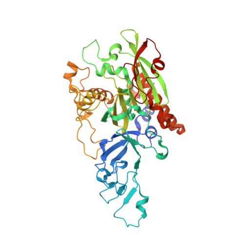
    Phthalate, a plasticizer, endocrine disruptor, and potential carcinogen, is degraded by a variety of bacteria. This degradation is initiated by phthalate dioxygenase (PDO), a Rieske oxygenase (RO) that catalyzes the dihydroxylation of phthalate to a dihydrodiol. PDO has long served as a model for understanding ROs despite a lack of structural data. Here we purified PDO KF1 from Comamonas testosteroni KF1 and found that it had an apparent k cat /K m for phthalate of 0.58 ± 0.09 μM -1 s -1 , over 25-fold greater than for terephthalate. The crystal structure of the enzyme at 2.1 Å resolution revealed that it is a hexamer comprising two stacked α 3 trimers, a configuration not previously observed in RO crystal structures. We show that within each trimer, the protomers adopt a head-to-tail configuration typical of ROs. The stacking of the trimers is stabilized by two extended helices, which make the catalytic domain of PDO KF1 larger than that of other characterized ROs. Complexes of PDO KF1 with phthalate and terephthalate revealed that Arg207 and Arg244, two residues on one face of the active site, position these substrates for regiospecific hydroxylation. Consistent with their roles as determinants of substrate specificity, substitution of either residue with alanine yielded variants that did not detectably turnover phthalate. Together, these results provide critical insights into a pollutant-degrading enzyme that has served as a paradigm for ROs and facilitate the engineering of this enzyme for bioremediation and biocatalytic applications.


  • Organizational Affiliation

    Department of Biosciences and Bioengineering, IIT Roorkee, Roorkee, India.


Macromolecules
Find similar proteins by:  (by identity cutoff)  |  3D Structure
Entity ID: 1
MoleculeChains Sequence LengthOrganismDetailsImage
Putative Phthalate 4,5-dioxygenase, subunit alpha441Cupriavidus metallidurans CH34Mutation(s): 0 
Gene Names: Rmet_5548
EC: 1.14.12.7
UniProt
Find proteins for Q1LBR9 (Cupriavidus metallidurans (strain ATCC 43123 / DSM 2839 / NBRC 102507 / CH34))
Explore Q1LBR9 
Go to UniProtKB:  Q1LBR9
Entity Groups  
Sequence Clusters30% Identity50% Identity70% Identity90% Identity95% Identity100% Identity
UniProt GroupQ1LBR9
Sequence Annotations
Expand
  • Reference Sequence
Small Molecules
Ligands 6 Unique
IDChains Name / Formula / InChI Key2D Diagram3D Interactions
FES (Subject of Investigation/LOI)
Query on FES

Download Ideal Coordinates CCD File 
G [auth A]FE2/S2 (INORGANIC) CLUSTER
Fe2 S2
NIXDOXVAJZFRNF-UHFFFAOYSA-N
GLU
Query on GLU

Download Ideal Coordinates CCD File 
E [auth A]GLUTAMIC ACID
C5 H9 N O4
WHUUTDBJXJRKMK-VKHMYHEASA-N
GOL
Query on GOL

Download Ideal Coordinates CCD File 
J [auth A]GLYCEROL
C3 H8 O3
PEDCQBHIVMGVHV-UHFFFAOYSA-N
GLY
Query on GLY

Download Ideal Coordinates CCD File 
F [auth A]GLYCINE
C2 H5 N O2
DHMQDGOQFOQNFH-UHFFFAOYSA-N
EDO
Query on EDO

Download Ideal Coordinates CCD File 
B [auth A],
C [auth A],
D [auth A],
I [auth A]
1,2-ETHANEDIOL
C2 H6 O2
LYCAIKOWRPUZTN-UHFFFAOYSA-N
FE2 (Subject of Investigation/LOI)
Query on FE2

Download Ideal Coordinates CCD File 
H [auth A]FE (II) ION
Fe
CWYNVVGOOAEACU-UHFFFAOYSA-N
Experimental Data & Validation

Experimental Data

  • Method: X-RAY DIFFRACTION
  • Resolution: 1.84 Å
  • R-Value Free: 0.212 
  • R-Value Work: 0.164 
  • R-Value Observed: 0.167 
  • Space Group: P 63
Unit Cell:
Length ( Å )Angle ( ˚ )
a = 103.416α = 90
b = 103.416β = 90
c = 77.59γ = 120
Software Package:
Software NamePurpose
REFMACrefinement
PDB_EXTRACTdata extraction
DIALSdata reduction
Aimlessdata scaling
Auto-Rickshawphasing

Structure Validation

View Full Validation Report


Entry History & Funding Information

Deposition Data


Funding OrganizationLocationGrant Number
Department of Biotechnology (DBT, India)IndiaBT/HRD/NBA/37/01/2015 (VIII)

Revision History  (Full details and data files)

  • Version 1.0: 2021-12-15
    Type: Initial release
  • Version 1.1: 2022-02-16
    Changes: Database references