7F79

Crystal structure of glutamate dehydrogenase 3 from Candida albicans in complex with alpha-ketoglutarate and NADPH


Experimental Data Snapshot

  • Method: X-RAY DIFFRACTION
  • Resolution: 2.70 Å
  • R-Value Free: 0.247 
  • R-Value Work: 0.199 
  • R-Value Observed: 0.201 

wwPDB Validation   3D Report Full Report


Ligand Structure Quality Assessment 


This is version 1.2 of the entry. See complete history


Literature

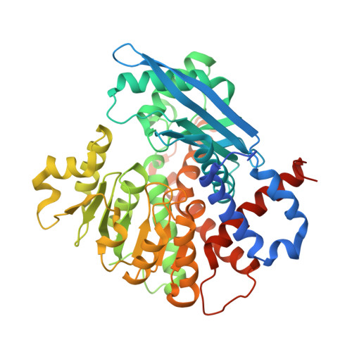
Crystal structure of glutamate dehydrogenase 3 from Candida albicans.

Li, N.Wang, W.Zeng, X.Liu, M.Li, M.Li, C.Wang, M.

(2021) Biochem Biophys Res Commun 570: 15-20

  • DOI: https://doi.org/10.1016/j.bbrc.2021.07.014
  • Primary Citation of Related Structures:  
    7F77, 7F79

  • PubMed Abstract: 

    Glutamate dehydrogenase 3 from Candida albicans (CaGdh3) catalyzes the reversible oxidative deamination of l-glutamate, playing an important role in the yeast-to-hyphal transition of C. albicans. Here we report the crystal structures of CaGdh3 and its complex with α-ketoglutarate and NADPH. CaGdh3 exists as a hexamer, with each subunit containing two domains. The substrate and coenzyme bind in the cleft between the two domains and their binding induces a conformational change in CaGdh3. Our results will help to understand the catalytic mechanism of CaGdh3 and will provide a structural basis for the design of antifungal drugs targeting the CaGdh3 pathway.


  • Organizational Affiliation

    Institutes of Physical Science and Information Technology, Anhui University, Hefei, 230601, Anhui, China.


Macromolecules
Find similar proteins by:  (by identity cutoff)  |  3D Structure
Entity ID: 1
MoleculeChains Sequence LengthOrganismDetailsImage
Glutamate dehydrogenase
A, B, C, D, E
A, B, C, D, E, F
484Candida albicans SC5314Mutation(s): 0 
Gene Names: GDH3
UniProt
Find proteins for A0A1D8PMH8 (Candida albicans (strain SC5314 / ATCC MYA-2876))
Explore A0A1D8PMH8 
Go to UniProtKB:  A0A1D8PMH8
Entity Groups  
Sequence Clusters30% Identity50% Identity70% Identity90% Identity95% Identity100% Identity
UniProt GroupA0A1D8PMH8
Sequence Annotations
Expand
  • Reference Sequence
Small Molecules
Ligands 3 Unique
IDChains Name / Formula / InChI Key2D Diagram3D Interactions
NDP (Subject of Investigation/LOI)
Query on NDP

Download Ideal Coordinates CCD File 
G [auth A]
J [auth B]
M [auth C]
O [auth D]
S [auth E]
G [auth A],
J [auth B],
M [auth C],
O [auth D],
S [auth E],
V [auth F]
NADPH DIHYDRO-NICOTINAMIDE-ADENINE-DINUCLEOTIDE PHOSPHATE
C21 H30 N7 O17 P3
ACFIXJIJDZMPPO-NNYOXOHSSA-N
AKG (Subject of Investigation/LOI)
Query on AKG

Download Ideal Coordinates CCD File 
H [auth A]
K [auth B]
N [auth C]
P [auth D]
T [auth E]
H [auth A],
K [auth B],
N [auth C],
P [auth D],
T [auth E],
W [auth F]
2-OXOGLUTARIC ACID
C5 H6 O5
KPGXRSRHYNQIFN-UHFFFAOYSA-N
GOL
Query on GOL

Download Ideal Coordinates CCD File 
I [auth A],
L [auth B],
Q [auth D],
R [auth D],
U [auth E]
GLYCEROL
C3 H8 O3
PEDCQBHIVMGVHV-UHFFFAOYSA-N
Experimental Data & Validation

Experimental Data

  • Method: X-RAY DIFFRACTION
  • Resolution: 2.70 Å
  • R-Value Free: 0.247 
  • R-Value Work: 0.199 
  • R-Value Observed: 0.201 
  • Space Group: P 1 21 1
Unit Cell:
Length ( Å )Angle ( ˚ )
a = 96.279α = 90
b = 155.224β = 95.5
c = 98.807γ = 90
Software Package:
Software NamePurpose
HKL-2000data reduction
HKL-2000data scaling
PHENIXrefinement
PDB_EXTRACTdata extraction
MoRDaphasing

Structure Validation

View Full Validation Report


Ligand Structure Quality Assessment 


Entry History & Funding Information

Deposition Data


Funding OrganizationLocationGrant Number
Other governmentChina1808085MC53

Revision History  (Full details and data files)

  • Version 1.0: 2021-07-21
    Type: Initial release
  • Version 1.1: 2021-07-28
    Changes: Database references
  • Version 1.2: 2023-11-29
    Changes: Data collection, Database references, Refinement description