


Structural variability of Mycobacterium tuberculosis SSB and susceptibility to inhibition.
Srikalaivani, R., Paul, A., Sriram, R., Narayanan, S., Shee, S., Singh, A., Varshney, U., Gopal, B., Vijayan, M.(2022) Curr Sci 122: 281-289
Experimental Data Snapshot
Starting Model: experimental
View more details
wwPDB Validation 3D Report Full Report

(2022) Curr Sci 122: 281-289
Entity ID: 1 | |||||
|---|---|---|---|---|---|
| Molecule | Chains | Sequence Length | Organism | Details | Image |
| Single-stranded DNA-binding protein | 164 | Mycobacterium tuberculosis H37Rv | Mutation(s): 0 Gene Names: ssb, Rv0054, MTCY21D4.17 | 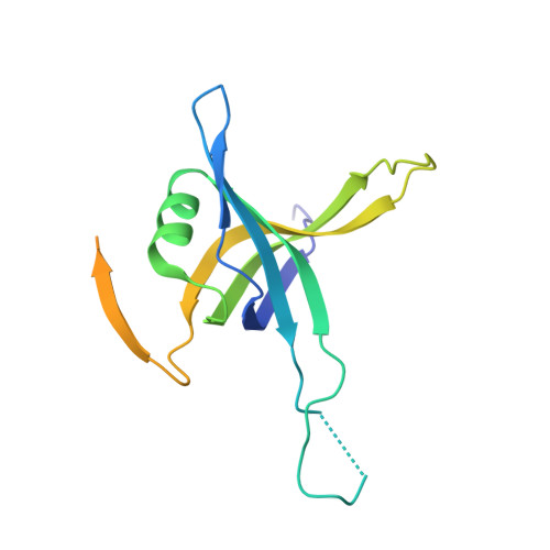 | |
UniProt | |||||
Entity Groups | |||||
| Sequence Clusters | 30% Identity50% Identity70% Identity90% Identity95% Identity100% Identity | ||||
| UniProt Group | P9WGD5 | ||||
Sequence AnnotationsExpand | |||||
Reference Sequence | |||||
| Length ( Å ) | Angle ( ˚ ) |
|---|---|
| a = 110.04 | α = 90 |
| b = 110.04 | β = 90 |
| c = 104.12 | γ = 120 |
| Software Name | Purpose |
|---|---|
| REFMAC | refinement |
| MOSFLM | data reduction |
| SCALA | data scaling |
| PHASER | phasing |
| Funding Organization | Location | Grant Number |
|---|---|---|
| Office of the Principal Scientific Adviser to the Government of India | India | -- |


