7F4C

The crystal structure of the immature holo-enzyme of homoserine dehydrogenase complexed with NADP and 1,4-butandiol from the hyperthermophilic archaeon Sulfurisphaera tokodaii.


Experimental Data Snapshot

  • Method: X-RAY DIFFRACTION
  • Resolution: 1.90 Å
  • R-Value Free: 0.257 
  • R-Value Work: 0.211 
  • R-Value Observed: 0.213 

wwPDB Validation   3D Report Full Report


Ligand Structure Quality Assessment 


This is version 1.2 of the entry. See complete history


Literature

Conformational changes in the catalytic region are responsible for heat-induced activation of hyperthermophilic homoserine dehydrogenase.

Kubota, T.Kurihara, E.Watanabe, K.Ogata, K.Kaneko, R.Goto, M.Ohshima, T.Yoshimune, K.

(2022) Commun Biol 5: 704-704

  • DOI: https://doi.org/10.1038/s42003-022-03656-7
  • Primary Citation of Related Structures:  
    7F4B, 7F4C

  • PubMed Abstract: 

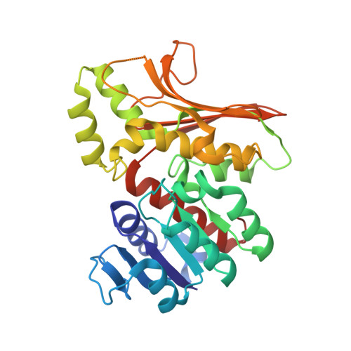
    When overexpressed as an immature enzyme in the mesophilic bacterium Escherichia coli, recombinant homoserine dehydrogenase from the hyperthermophilic archaeon Sulfurisphaera tokodaii (StHSD) was markedly activated by heat treatment. Both the apo- and holo-forms of the immature enzyme were successively crystallized, and the two structures were determined. Comparison among the structures of the immature enzyme and previously reported structures of mature enzymes revealed that a conformational change in a flexible part (residues 160-190) of the enzyme, which encloses substrates within the substrate-binding pocket, is smaller in the immature enzyme. The immature enzyme, but not the mature enzyme, formed a complex that included NADP + , despite its absence during crystallization. This indicates that the opening to the substrate-binding pocket in the immature enzyme is not sufficient for substrate-binding, efficient catalytic turnover or release of NADP + . Thus, specific conformational changes within the catalytic region appear to be responsible for heat-induced activation.


  • Organizational Affiliation

    Department of Applied Molecular Chemistry, Graduate School of Industrial Technology, Nihon University, 1-2-1, Izumichou, Narashino, Chiba, 275-8575, Japan.


Macromolecules
Find similar proteins by:  (by identity cutoff)  |  3D Structure
Entity ID: 1
MoleculeChains Sequence LengthOrganismDetailsImage
Homoserine dehydrogenase
A, B
304Sulfurisphaera tokodaii str. 7Mutation(s): 0 
Gene Names: homST1519STK_15190
EC: 1.1.1.3
UniProt
Find proteins for F9VNG5 (Sulfurisphaera tokodaii (strain DSM 16993 / JCM 10545 / NBRC 100140 / 7))
Explore F9VNG5 
Go to UniProtKB:  F9VNG5
Entity Groups  
Sequence Clusters30% Identity50% Identity70% Identity90% Identity95% Identity100% Identity
UniProt GroupF9VNG5
Sequence Annotations
Expand
  • Reference Sequence
Experimental Data & Validation

Experimental Data

  • Method: X-RAY DIFFRACTION
  • Resolution: 1.90 Å
  • R-Value Free: 0.257 
  • R-Value Work: 0.211 
  • R-Value Observed: 0.213 
  • Space Group: P 1 21 1
Unit Cell:
Length ( Å )Angle ( ˚ )
a = 55.328α = 90
b = 79.052β = 106.51
c = 65.914γ = 90
Software Package:
Software NamePurpose
SCALAdata scaling
REFMACrefinement
PDB_EXTRACTdata extraction
XDSdata reduction
MOLREPphasing

Structure Validation

View Full Validation Report


Ligand Structure Quality Assessment 


Entry History 

Deposition Data

Revision History  (Full details and data files)

  • Version 1.0: 2022-06-22
    Type: Initial release
  • Version 1.1: 2022-08-10
    Changes: Database references
  • Version 1.2: 2023-11-29
    Changes: Data collection, Refinement description