7F45 | pdb_00007f45

Structure of an Anti-CRISPR protein


Experimental Data Snapshot

  • Method: X-RAY DIFFRACTION
  • Resolution: 3.52 Å
  • R-Value Free: 
    0.311 (Depositor), 0.288 (DCC) 
  • R-Value Work: 
    0.255 (Depositor), 0.259 (DCC) 
  • R-Value Observed: 
    0.257 (Depositor) 

Starting Model: experimental
View more details

wwPDB Validation 3D Report Full Report

Validation slider image for 7F45

This is version 1.3 of the entry. See complete history

Literature

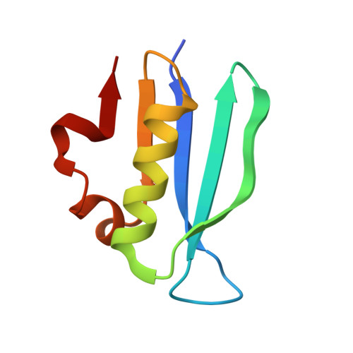
AcrIF5 specifically targets DNA-bound CRISPR-Cas surveillance complex for inhibition.

Xie, Y.Zhang, L.Gao, Z.Yin, P.Wang, H.Li, H.Chen, Z.Zhang, Y.Yang, M.Feng, Y.

(2022) Nat Chem Biol 18: 670-677

  • DOI: https://doi.org/10.1038/s41589-022-00995-8
  • Primary Citation Related Structures: 
    7EQG, 7F45

  • PubMed Abstract: 

    CRISPR-Cas systems are prokaryotic antiviral systems, and phages use anti-CRISPR proteins (Acrs) to inactivate these systems. Here we present structural and functional analyses of AcrIF5, exploring its unique anti-CRISPR mechanism. AcrIF5 shows binding specificity only for the target DNA-bound form of the crRNA-guided surveillance (Csy) complex, but not the apo Csy complex from the type I-F CRISPR-Cas system. We solved the structure of the Csy-dsDNA-AcrIF5 complex, revealing that the conformational changes of the Csy complex caused by dsDNA binding dictate the binding specificity for the Csy-dsDNA complex by AcrIF5. Mechanistically, five AcrIF5 molecules bind one Csy-dsDNA complex, which destabilizes the helical bundle domain of Cas8f, thus preventing subsequent Cas2/3 recruitment. AcrIF5 exists in symbiosis with AcrIF3, which blocks Cas2/3 recruitment. This attack on the recruitment event stands in contrast to the conventional mechanisms of blocking binding of target DNA. Overall, our study reveals an unprecedented mechanism of CRISPR-Cas inhibition by AcrIF5.


  • Organizational Affiliation
    • Beijing Advanced Innovation Center for Soft Matter Science and Engineering, Beijing Key Laboratory of Bioprocess, State Key Laboratory of Chemical Resource Engineering, College of Life Science and Technology, Beijing University of Chemical Technology, Beijing, China.

Macromolecule Content 

  • Total Structure Weight: 17.06 kDa 
  • Atom Count: 1,107 
  • Modeled Residue Count: 152 
  • Deposited Residue Count: 158 
  • Unique protein chains: 1

Macromolecules

Find similar proteins by:|  3D Structure
Entity ID: 1
MoleculeChains  Sequence LengthOrganismDetailsImage
AcrIF5A,
B [auth P]
79Pseudomonas aeruginosaMutation(s): 0 
Gene Names: IPC1164_31450

Experimental Data & Validation

Experimental Data

  • Method: X-RAY DIFFRACTION
  • Resolution: 3.52 Å
  • R-Value Free:  0.311 (Depositor), 0.288 (DCC) 
  • R-Value Work:  0.255 (Depositor), 0.259 (DCC) 
  • R-Value Observed: 0.257 (Depositor) 
Space Group: P 41 21 2
Unit Cell:
Length ( Å )Angle ( ˚ )
a = 66.741α = 90
b = 66.741β = 90
c = 120.98γ = 90
Software Package:
Software NamePurpose
SCALEPACKdata scaling
PHENIXrefinement
PDB_EXTRACTdata extraction
SCALEPACKdata scaling
PHASERphasing
HKL-2000data reduction

Structure Validation

View Full Validation Report


Entry History 

& Funding Information

Deposition Data

  • Released Date: 2022-03-09 
  • Deposition Author(s): Feng, Y.

Funding OrganizationLocationGrant Number
National Science Foundation (NSF, China)China31822012

Revision History  (Full details and data files)

  • Version 1.0: 2022-03-09
    Type: Initial release
  • Version 1.1: 2022-04-06
    Changes: Database references
  • Version 1.2: 2022-06-08
    Changes: Database references
  • Version 1.3: 2023-11-29
    Changes: Data collection, Refinement description