


Directed evolution of cytochrome P450DA hydroxylase activity for stereoselective biohydroxylation
Wan, N.W., Cui, H.B., Zhao, L., Shan, J., Chen, K., Wang, Z.Q., Zhou, X.J., Cui, B.D., Han, W.Y., Chen, Y.Z.(2022) Catal Sci Technol 12: 5703-5708
Experimental Data Snapshot
Starting Model: experimental
View more details
(2022) Catal Sci Technol 12: 5703-5708
Entity ID: 1 | |||||
|---|---|---|---|---|---|
| Molecule | Chains | Sequence Length | Organism | Details | Image |
| Bifunctional cytochrome P450/NADPH--P450 reductase | 501 | Deinococcus aerius | Mutation(s): 0 Gene Names: DAERI_140002 EC: 1.14.14.1 (PDB Primary Data), 1.6.2.4 (PDB Primary Data) | 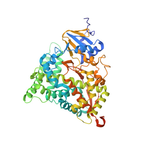 | |
UniProt | |||||
Entity Groups | |||||
| Sequence Clusters | 30% Identity50% Identity70% Identity90% Identity95% Identity100% Identity | ||||
| UniProt Group | A0A2I9DQ46 | ||||
Sequence AnnotationsExpand | |||||
Reference Sequence | |||||
| Ligands 3 Unique | |||||
|---|---|---|---|---|---|
| ID | Chains | Name / Formula / InChI Key | 2D Diagram | 3D Interactions | |
| HEM (Subject of Investigation/LOI) Download:Ideal Coordinates CCD File | E [auth A], I [auth B], J [auth C], K [auth D] | PROTOPORPHYRIN IX CONTAINING FE C34 H32 Fe N4 O4 KABFMIBPWCXCRK-RGGAHWMASA-L |  | ||
| SER Download:Ideal Coordinates CCD File | H [auth B] | SERINE C3 H7 N O3 MTCFGRXMJLQNBG-REOHCLBHSA-N |  | ||
| SO4 Download:Ideal Coordinates CCD File | F [auth A], G [auth A], L [auth D] | SULFATE ION O4 S QAOWNCQODCNURD-UHFFFAOYSA-L |  | ||
| Length ( Å ) | Angle ( ˚ ) |
|---|---|
| a = 426.301 | α = 90 |
| b = 62.799 | β = 90 |
| c = 95.188 | γ = 90 |
| Software Name | Purpose |
|---|---|
| PHENIX | refinement |
| HKL-3000 | data reduction |
| HKL-3000 | data scaling |
| PHASER | phasing |


