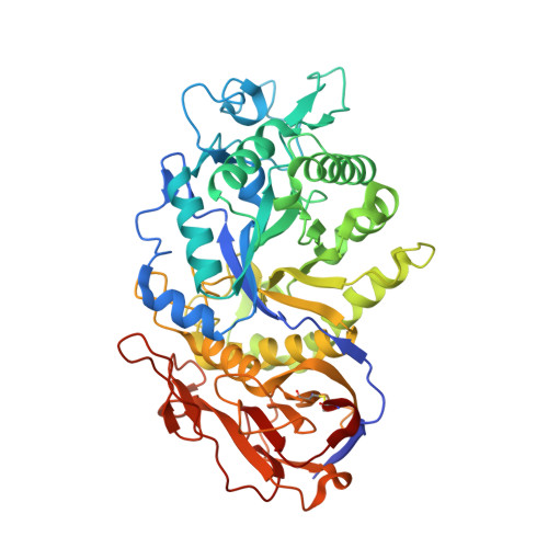
7EYO

Crystal structure of leech hyaluronidase


Experimental Data Snapshot

  • Method: X-RAY DIFFRACTION
  • Resolution: 1.85 Å
  • R-Value Free: 0.212 
  • R-Value Work: 0.177 
  • R-Value Observed: 0.179 

Starting Model: experimental
View more details

wwPDB Validation   3D Report Full Report


This is version 1.1 of the entry. See complete history


Literature

Structure and cleavage pattern of a hyaluronate 3-glycanohydrolase in the glycoside hydrolase 79 family.

Huang, H.Hou, X.Xu, R.Deng, Z.Wang, Y.Du, G.Rao, Y.Chen, J.Kang, Z.

(2022) Carbohydr Polym 277: 118838-118838

  • DOI: https://doi.org/10.1016/j.carbpol.2021.118838
  • Primary Citation of Related Structures:  
    7EYO

  • PubMed Abstract: 

    Hyaluronidases have attracted a great deal of interest in the field of medicine due to their fundamental roles in the breakdown of hyaluronan. However, little is known about the catalytic mechanism of the hyaluronate 3-glycanohydrolases. Here, we report the crystal structure and cleavage pattern of a leech hyaluronidase (LHyal), which hydrolyzes the β-1,3-glycosidic bonds of hyaluronan. LHyal exhibits the typical structural features of glycoside hydrolase 79 family but contains a variable 'exo-pocket' loop where basic residues R102 and K103 are the structural determinants of hyaluronan binding. Through analysis of the hydrolysis of even- and odd-numbered hyaluronan oligosaccharides, we demonstrate that hexasaccharide is the shortest natural substrate, which can be cleaved from both the reducing and non-reducing ends to release disaccharides, and pentasaccharides are the smallest fragments for recognition and hydrolysis. These observations provide new insights into the degradation of hyaluronan and the evolutionary relationships of the GH79 family enzymes.


  • Organizational Affiliation

    The Key Laboratory of Carbohydrate Chemistry and Biotechnology, Ministry of Education, School of Biotechnology, Jiangnan University, Wuxi 214122, China; The Science Center for Future Foods, Jiangnan University, Wuxi 214122, China.


Macromolecules
Find similar proteins by:  (by identity cutoff)  |  3D Structure
Entity ID: 1
MoleculeChains Sequence LengthOrganismDetailsImage
Hyaluronoglucuronidase496Hirudo nipponiaMutation(s): 0 
EC: 3.2.1.36
UniProt
Find proteins for X4Y2L4 (Hirudo nipponia)
Explore X4Y2L4 
Go to UniProtKB:  X4Y2L4
Entity Groups  
Sequence Clusters30% Identity50% Identity70% Identity90% Identity95% Identity100% Identity
UniProt GroupX4Y2L4
Glycosylation
Glycosylation Sites: 2
Sequence Annotations
Expand
  • Reference Sequence
Oligosaccharides

Help

Entity ID: 2
MoleculeChains Length2D Diagram Glycosylation3D Interactions
2-acetamido-2-deoxy-beta-D-glucopyranose-(1-4)-2-acetamido-2-deoxy-beta-D-glucopyranose
B, C
2N-Glycosylation
Glycosylation Resources
GlyTouCan:  G42666HT
GlyCosmos:  G42666HT
GlyGen:  G42666HT
Experimental Data & Validation

Experimental Data

  • Method: X-RAY DIFFRACTION
  • Resolution: 1.85 Å
  • R-Value Free: 0.212 
  • R-Value Work: 0.177 
  • R-Value Observed: 0.179 
  • Space Group: P 1 21 1
Unit Cell:
Length ( Å )Angle ( ˚ )
a = 43.514α = 90
b = 69.474β = 95.97
c = 86.739γ = 90
Software Package:
Software NamePurpose
HKL-2000data scaling
REFMACrefinement
PDB_EXTRACTdata extraction
HKL-2000data reduction
MOLREPphasing

Structure Validation

View Full Validation Report


Entry History & Funding Information

Deposition Data


Funding OrganizationLocationGrant Number
National Natural Science Foundation of China (NSFC)China32000058
National Natural Science Foundation of China (NSFC)China31970085
Other privateChinaBK20200025
Other privateChina2018YFA0901400

Revision History  (Full details and data files)

  • Version 1.0: 2022-05-25
    Type: Initial release
  • Version 1.1: 2023-11-29
    Changes: Data collection, Refinement description