7EXO

Structure of legume lectin domain from Methanocaldococcus jannaschii in mannose bound form


Experimental Data Snapshot

  • Method: X-RAY DIFFRACTION
  • Resolution: 1.75 Å
  • R-Value Free: 0.183 
  • R-Value Work: 0.159 

wwPDB Validation   3D Report Full Report


Ligand Structure Quality Assessment 


This is version 1.4 of the entry. See complete history


Literature

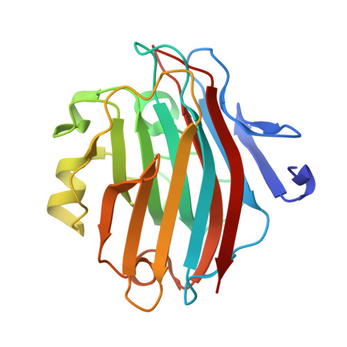
Crystal structure of an L-type lectin domain from archaea.

Khan, F.Kaza, S.

(2023) Proteins 91: 456-465

  • DOI: https://doi.org/10.1002/prot.26440
  • Primary Citation of Related Structures:  
    7ELV, 7EXO

  • PubMed Abstract: 

    The crystal structures of an L-type lectin domain from Methanocaldococcus jannaschii in apo and mannose-bound forms have been determined. A thorough investigation of L-type lectin domains from several organisms provides insight into the differences in these domains from different kingdoms of life. While the overall fold of the L-type lectin domain is conserved, differences in the lengths of the carbohydrate-binding loops and significant variations in the Mn 2+ -binding site compared to the Ca 2+ -binding site are observed. Furthermore, the sequence and phylogenetic analyses suggest that the archaeal L-type lectin domain is evolutionarily closer to the plant legume lectins than to its bacterial or animal counterparts. This is the first report of the biochemical, structural, sequence, and phylogenetic analyses of an L-type lectin domain from archaea and serves to enhance our understanding of the species-specific differences and evolution of L-type lectin domains.


  • Organizational Affiliation

    Molecular Biophysics Unit, Indian Institute of Science, Bangalore, Karnataka, India.


Macromolecules
Find similar proteins by:  (by identity cutoff)  |  3D Structure
Entity ID: 1
MoleculeChains Sequence LengthOrganismDetailsImage
Legume lectin209Methanocaldococcus jannaschii DSM 2661Mutation(s): 0 
Gene Names: MJ1396
UniProt
Find proteins for Q58791 (Methanocaldococcus jannaschii (strain ATCC 43067 / DSM 2661 / JAL-1 / JCM 10045 / NBRC 100440))
Explore Q58791 
Go to UniProtKB:  Q58791
Entity Groups  
Sequence Clusters30% Identity50% Identity70% Identity90% Identity95% Identity100% Identity
UniProt GroupQ58791
Sequence Annotations
Expand
  • Reference Sequence
Small Molecules
Ligands 6 Unique
IDChains Name / Formula / InChI Key2D Diagram3D Interactions
BMA (Subject of Investigation/LOI)
Query on BMA

Download Ideal Coordinates CCD File 
C [auth A]beta-D-mannopyranose
C6 H12 O6
WQZGKKKJIJFFOK-RWOPYEJCSA-N
MPD
Query on MPD

Download Ideal Coordinates CCD File 
B [auth A],
E [auth A]
(4S)-2-METHYL-2,4-PENTANEDIOL
C6 H14 O2
SVTBMSDMJJWYQN-YFKPBYRVSA-N
GOL
Query on GOL

Download Ideal Coordinates CCD File 
F [auth A],
G [auth A]
GLYCEROL
C3 H8 O3
PEDCQBHIVMGVHV-UHFFFAOYSA-N
ACT
Query on ACT

Download Ideal Coordinates CCD File 
D [auth A]ACETATE ION
C2 H3 O2
QTBSBXVTEAMEQO-UHFFFAOYSA-M
MN
Query on MN

Download Ideal Coordinates CCD File 
I [auth A]MANGANESE (II) ION
Mn
WAEMQWOKJMHJLA-UHFFFAOYSA-N
CA
Query on CA

Download Ideal Coordinates CCD File 
H [auth A]CALCIUM ION
Ca
BHPQYMZQTOCNFJ-UHFFFAOYSA-N
Experimental Data & Validation

Experimental Data

  • Method: X-RAY DIFFRACTION
  • Resolution: 1.75 Å
  • R-Value Free: 0.183 
  • R-Value Work: 0.159 
  • Space Group: P 41 21 2
Unit Cell:
Length ( Å )Angle ( ˚ )
a = 55.03α = 90
b = 55.03β = 90
c = 149.05γ = 90
Software Package:
Software NamePurpose
REFMACrefinement
iMOSFLMdata reduction
Aimlessdata scaling
PHASERphasing

Structure Validation

View Full Validation Report


Ligand Structure Quality Assessment 


Entry History 

Deposition Data

Revision History  (Full details and data files)

  • Version 1.0: 2022-06-01
    Type: Initial release
  • Version 1.1: 2022-12-14
    Changes: Database references, Derived calculations
  • Version 1.2: 2022-12-21
    Changes: Database references
  • Version 1.3: 2023-03-22
    Changes: Database references
  • Version 1.4: 2023-11-29
    Changes: Data collection, Refinement description