7EU8 | pdb_00007eu8

Structure of the human GluN1-GluN2B NMDA receptor in complex with S-ketamine,glycine and glutamate

  • Classification: MEMBRANE PROTEIN
  • Organism(s): Homo sapiens
  • Expression System: Homo sapiens
  • Mutation(s): No 
  • Membrane Protein: Yes  OPMPDBTMmpstruc

  • Deposited: 2021-05-16 Released: 2021-07-28 
  • Deposition Author(s): Zhang, T., Zhang, Y., Zhu, S.
  • Funding Organization(s): National Natural Science Foundation of China (NSFC), Chinese Academy of Sciences, National Key R&D Program of China, Shanghai Municipal Health Commission in China, Shanghai Municipal Science and Technology Major Project

Experimental Data Snapshot

  • Method: ELECTRON MICROSCOPY
  • Resolution: 4.07 Å
  • Aggregation State: PARTICLE 
  • Reconstruction Method: SINGLE PARTICLE 

wwPDB Validation 3D Report Full Report

Validation slider image for 7EU8

This is version 1.4 of the entry. See complete history

Literature

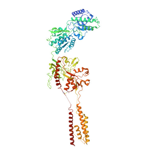
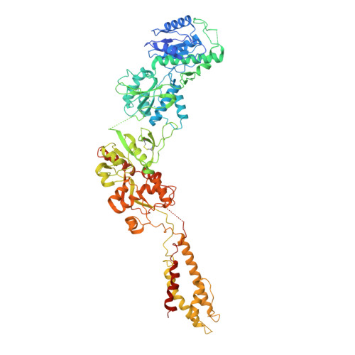
Structural basis of ketamine action on human NMDA receptors.

Zhang, Y.Ye, F.Zhang, T.Lv, S.Zhou, L.Du, D.Lin, H.Guo, F.Luo, C.Zhu, S.

(2021) Nature 596: 301-305

  • DOI: https://doi.org/10.1038/s41586-021-03769-9
  • Primary Citation Related Structures: 
    7EU7, 7EU8

  • PubMed Abstract: 

    Ketamine is a non-competitive channel blocker of N-methyl-D-aspartate (NMDA) receptors 1 . A single sub-anaesthetic dose of ketamine produces rapid (within hours) and long-lasting antidepressant effects in patients who are resistant to other antidepressants 2,3 . Ketamine is a racemic mixture of S- and R-ketamine enantiomers, with S-ketamine isomer being the more active antidepressant 4 . Here we describe the cryo-electron microscope structures of human GluN1-GluN2A and GluN1-GluN2B NMDA receptors in complex with S-ketamine, glycine and glutamate. Both electron density maps uncovered the binding pocket for S-ketamine in the central vestibule between the channel gate and selectivity filter. Molecular dynamics simulation showed that S-ketamine moves between two distinct locations within the binding pocket. Two amino acids-leucine 642 on GluN2A (homologous to leucine 643 on GluN2B) and asparagine 616 on GluN1-were identified as key residues that form hydrophobic and hydrogen-bond interactions with ketamine, and mutations at these residues reduced the potency of ketamine in blocking NMDA receptor channel activity. These findings show structurally how ketamine binds to and acts on human NMDA receptors, and pave the way for the future development of ketamine-based antidepressants.


  • Organizational Affiliation
    • Institute of Neuroscience, State Key Laboratory of Neuroscience, CAS Center for Excellence in Brain Science and Intelligence Technology, Chinese Academy of Sciences, Shanghai, China.

Macromolecule Content 

  • Total Structure Weight: 385.41 kDa 
  • Atom Count: 18,670 
  • Modeled Residue Count: 3,010 
  • Deposited Residue Count: 3,418 
  • Unique protein chains: 2

Macromolecules

Find similar proteins by:|  3D Structure
Entity ID: 1
MoleculeChains  Sequence LengthOrganismDetailsImage
Glutamate receptor ionotropic, NMDA 1
A, C
847Homo sapiensMutation(s): 0 
Gene Names: GRIN1NMDAR1
Membrane Entity: Yes 
UniProt & NIH Common Fund Data Resources
Find proteins for Q05586 (Homo sapiens)
Explore Q05586 
Go to UniProtKB:  Q05586
PHAROS:  Q05586
GTEx:  ENSG00000176884 
Entity Groups
Sequence Clusters30% Identity50% Identity70% Identity90% Identity95% Identity100% Identity
UniProt GroupQ05586
Glycosylation
Glycosylation Sites: 1Go to GlyGen: Q05586-1
Sequence Annotations
Expand
Reference Sequence
Find similar proteins by:|  3D Structure
Entity ID: 2
MoleculeChains  Sequence LengthOrganismDetailsImage
Glutamate receptor ionotropic, NMDA 2B
B, D
862Homo sapiensMutation(s): 0 
Gene Names: GRIN2B
Membrane Entity: Yes 
UniProt & NIH Common Fund Data Resources
Find proteins for Q13224 (Homo sapiens)
Explore Q13224 
Go to UniProtKB:  Q13224
PHAROS:  Q13224
GTEx:  ENSG00000273079 
Entity Groups
Sequence Clusters30% Identity50% Identity70% Identity90% Identity95% Identity100% Identity
UniProt GroupQ13224
Glycosylation
Glycosylation Sites: 3Go to GlyGen: Q13224-1
Sequence Annotations
Expand
Reference Sequence

Small Molecules

Ligands 2 Unique
IDChains Name / Formula / InChI Key2D Diagram3D Interactions
JC9
(Subject of Investigation/LOI)

Query on JC9


Download:Ideal Coordinates CCD File
H [auth B](2~{S})-2-(2-chlorophenyl)-2-(methylamino)cyclohexan-1-one
C13 H16 Cl N O
YQEZLKZALYSWHR-ZDUSSCGKSA-N
NAG

Query on NAG


Download:Ideal Coordinates CCD File
E [auth A]
F [auth B]
G [auth B]
I [auth C]
J [auth D]
E [auth A],
F [auth B],
G [auth B],
I [auth C],
J [auth D],
K [auth D],
L [auth D]
2-acetamido-2-deoxy-beta-D-glucopyranose
C8 H15 N O6
OVRNDRQMDRJTHS-FMDGEEDCSA-N

Experimental Data & Validation

Experimental Data

  • Method: ELECTRON MICROSCOPY
  • Resolution: 4.07 Å
  • Aggregation State: PARTICLE 
  • Reconstruction Method: SINGLE PARTICLE 

Structure Validation

View Full Validation Report


Entry History 

& Funding Information

Deposition Data


Funding OrganizationLocationGrant Number
National Natural Science Foundation of China (NSFC)China31771115
Chinese Academy of SciencesChinaXDBS01020000
National Natural Science Foundation of China (NSFC)China81625022
National Natural Science Foundation of China (NSFC)China91853205
National Natural Science Foundation of China (NSFC)China81821005
National Key R&D Program of ChinaChina2017YFA0505700
Shanghai Municipal Health Commission in ChinaChina18431907100
Shanghai Municipal Health Commission in ChinaChina19XD1404700
Shanghai Municipal Science and Technology Major ProjectChina2018SHZDZX05

Revision History  (Full details and data files)

  • Version 1.0: 2021-07-28
    Type: Initial release
  • Version 1.1: 2021-08-04
    Changes: Author supporting evidence, Database references, Structure summary
  • Version 1.2: 2021-09-22
    Changes: Database references, Structure summary
  • Version 1.3: 2022-10-26
    Changes: Structure summary
  • Version 1.4: 2024-11-06
    Changes: Data collection, Structure summary