


Structure-based interface engineering methodology in designing a thermostable amylose-forming transglucosylase
Tian, Y., Hou, X., Ni, D., Xu, W., Guang, C., Zhang, W., Chen, Q., Rao, Y., Mu, W.(2022) J Biological Chem 298: 102074
Experimental Data Snapshot
Starting Model: experimental
View more details
wwPDB Validation 3D Report Full Report

Entity ID: 1 | |||||
|---|---|---|---|---|---|
| Molecule | Chains | Sequence Length | Organism | Details | Image |
| amylosucrase | 656 | Calidithermus timidus DSM 17022 | Mutation(s): 0 EC: 2.4.1.4 | 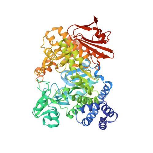 | |
UniProt | |||||
Find proteins for A0A915Q9D7 (Calidithermus timidus DSM 17022) Explore A0A915Q9D7 Go to UniProtKB: A0A915Q9D7 | |||||
Entity Groups | |||||
| Sequence Clusters | 30% Identity50% Identity70% Identity90% Identity95% Identity100% Identity | ||||
| UniProt Group | A0A915Q9D7 | ||||
Sequence AnnotationsExpand | |||||
Reference Sequence | |||||
| Ligands 1 Unique | |||||
|---|---|---|---|---|---|
| ID | Chains | Name / Formula / InChI Key | 2D Diagram | 3D Interactions | |
| TRS Download:Ideal Coordinates CCD File | E [auth A], F [auth B], G [auth C], H [auth D] | 2-AMINO-2-HYDROXYMETHYL-PROPANE-1,3-DIOL C4 H12 N O3 LENZDBCJOHFCAS-UHFFFAOYSA-O |  | ||
| Length ( Å ) | Angle ( ˚ ) |
|---|---|
| a = 105.75 | α = 90 |
| b = 126.66 | β = 90 |
| c = 294.73 | γ = 90 |
| Software Name | Purpose |
|---|---|
| REFMAC | refinement |
| HKL-2000 | data scaling |
| PDB_EXTRACT | data extraction |
| MOLREP | phasing |
| HEK-2000 | data reduction |
| Funding Organization | Location | Grant Number |
|---|---|---|
| National Natural Science Foundation of China (NSFC) | China | 31922073 |


