7EP3

Crystal structure of ZER1 bound to GAGN degron

  • Classification: LIGASE
  • Organism(s): Homo sapiens
  • Expression System: Escherichia coli
  • Mutation(s): No 

  • Deposited: 2021-04-26 Released: 2021-07-14 
  • Deposition Author(s): Yan, X., Li, Y.
  • Funding Organization(s): National Natural Science Foundation of China (NSFC)

Experimental Data Snapshot

  • Method: X-RAY DIFFRACTION
  • Resolution: 1.51 Å
  • R-Value Free: 0.256 
  • R-Value Work: 0.219 

wwPDB Validation   3D Report Full Report


This is version 1.2 of the entry. See complete history


Literature

Molecular basis for recognition of Gly/N-degrons by CRL2 ZYG11B and CRL2 ZER1 .

Yan, X.Li, Y.Wang, G.Zhou, Z.Song, G.Feng, Q.Zhao, Y.Mi, W.Ma, Z.Dong, C.

(2021) Mol Cell 81: 3262-3274.e3

  • DOI: https://doi.org/10.1016/j.molcel.2021.06.010
  • Primary Citation of Related Structures:  
    7EP0, 7EP1, 7EP2, 7EP3, 7EP4, 7EP5

  • PubMed Abstract: 

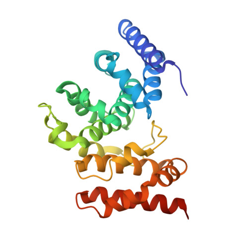
    N-degron pathways are a set of proteolytic systems that target the N-terminal destabilizing residues of substrates for proteasomal degradation. Recently, the Gly/N-degron pathway has been identified as a new branch of the N-degron pathway. The N-terminal glycine degron (Gly/N-degron) is recognized by ZYG11B and ZER1, the substrate receptors of the Cullin 2-RING E3 ubiquitin ligase (CRL2). Here we present the crystal structures of ZYG11B and ZER1 bound to various Gly/N-degrons. The structures reveal that ZYG11B and ZER1 utilize their armadillo (ARM) repeats forming a deep and narrow cavity to engage mainly the first four residues of Gly/N-degrons. The α-amino group of the Gly/N-degron is accommodated in an acidic pocket by five conserved hydrogen bonds. These structures, together with biochemical studies, decipher the molecular basis for the specific recognition of the Gly/N-degron by ZYG11B and ZER1, providing key information for future structure-based chemical probe design.


  • Organizational Affiliation

    Department of Biochemistry and Molecular Biology, The Province and Ministry Co-sponsored Collaborative Innovation Center for Medical Epigenetics, Key Laboratory of Immune Microenvironment and Disease (Ministry of Education), School of Basic Medical Sciences, Tianjin Medical University, Tianjin 300070, China.


Macromolecules
Find similar proteins by:  (by identity cutoff)  |  3D Structure
Entity ID: 1
MoleculeChains Sequence LengthOrganismDetailsImage
Protein zer-1 homolog251Homo sapiensMutation(s): 0 
Gene Names: ZER1C9orf60ZYGZYG11BL
UniProt & NIH Common Fund Data Resources
Find proteins for Q7Z7L7 (Homo sapiens)
Explore Q7Z7L7 
Go to UniProtKB:  Q7Z7L7
PHAROS:  Q7Z7L7
GTEx:  ENSG00000160445 
Entity Groups  
Sequence Clusters30% Identity50% Identity70% Identity90% Identity95% Identity100% Identity
UniProt GroupQ7Z7L7
Sequence Annotations
Expand
  • Reference Sequence
Experimental Data & Validation

Experimental Data

  • Method: X-RAY DIFFRACTION
  • Resolution: 1.51 Å
  • R-Value Free: 0.256 
  • R-Value Work: 0.219 
  • Space Group: C 2 2 21
Unit Cell:
Length ( Å )Angle ( ˚ )
a = 67.97α = 90
b = 120.42β = 90
c = 68γ = 90
Software Package:
Software NamePurpose
PHENIXrefinement
XDSdata reduction
XDSdata scaling
PHENIXphasing

Structure Validation

View Full Validation Report


Entry History & Funding Information

Deposition Data

  • Released Date: 2021-07-14 
  • Deposition Author(s): Yan, X., Li, Y.

Funding OrganizationLocationGrant Number
National Natural Science Foundation of China (NSFC)China31900865
National Natural Science Foundation of China (NSFC)China32071193
National Natural Science Foundation of China (NSFC)China81874039

Revision History  (Full details and data files)

  • Version 1.0: 2021-07-14
    Type: Initial release
  • Version 1.1: 2021-09-01
    Changes: Database references
  • Version 1.2: 2023-11-29
    Changes: Data collection, Refinement description