7EOQ

Structure of the human GluN1/GluN2A NMDA receptor in the glycine/CPP bound state

  • Classification: MEMBRANE PROTEIN
  • Organism(s): Homo sapiens
  • Expression System: Homo sapiens
  • Mutation(s): Yes 
  • Membrane Protein: Yes  OPMPDBTMMemProtMDmpstruc

  • Deposited: 2021-04-22 Released: 2021-06-30 
  • Deposition Author(s): Wang, H., Zhu, S.
  • Funding Organization(s): National Natural Science Foundation of China (NSFC), European Research Council (ERC), Strategic Priority Research Program of Chinese Academy of Sciences, Shanghai Municipal Science and Technology Major Project, National Key R&D Program of China

Experimental Data Snapshot

  • Method: ELECTRON MICROSCOPY
  • Resolution: 4.10 Å
  • Aggregation State: PARTICLE 
  • Reconstruction Method: SINGLE PARTICLE 

wwPDB Validation   3D Report Full Report


This is version 1.2 of the entry. See complete history


Literature

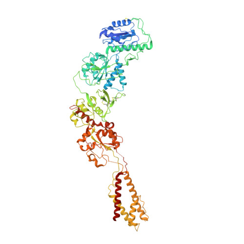
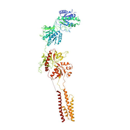
Gating mechanism and a modulatory niche of human GluN1-GluN2A NMDA receptors.

Wang, H.Lv, S.Stroebel, D.Zhang, J.Pan, Y.Huang, X.Zhang, X.Paoletti, P.Zhu, S.

(2021) Neuron 109: 2443-2456.e5

  • DOI: https://doi.org/10.1016/j.neuron.2021.05.031
  • Primary Citation of Related Structures:  
    7EOQ, 7EOR, 7EOS, 7EOT, 7EOU

  • PubMed Abstract: 

    N-methyl-D-aspartate (NMDA) receptors are glutamate-gated calcium-permeable ion channels that are widely implicated in synaptic transmission and plasticity. Here, we report a gallery of cryo-electron microscopy (cryo-EM) structures of the human GluN1-GluN2A NMDA receptor at an overall resolution of 4 Å in complex with distinct ligands or modulators. In the full-length context of GluN1-GluN2A receptors, we visualize the competitive antagonists bound to the ligand-binding domains (LBDs) of GluN1 and GluN2A subunits, respectively. We reveal that the binding of positive allosteric modulator shortens the distance between LBDs and the transmembrane domain (TMD), which further stretches the opening of the gate. In addition, we unexpectedly visualize the binding cavity of the "foot-in-the-door" blocker 9-aminoacridine within the LBD-TMD linker region, differing from the conventional "trapping" blocker binding site at the vestibule within the TMD. Our study provides molecular insights into the crosstalk between LBDs and TMD during channel activation, inhibition, and allosteric transition.


  • Organizational Affiliation

    Institute of Neuroscience, State Key Laboratory of Neuroscience, CAS Center for Excellence in Brain Science and Intelligence Technology, Chinese Academy of Sciences, Shanghai 200031, China; University of Chinese Academy of Sciences, Beijing 100049, China.


Macromolecules
Find similar proteins by:  (by identity cutoff)  |  3D Structure
Entity ID: 1
MoleculeChains Sequence LengthOrganismDetailsImage
Glutamate receptor ionotropic, NMDA 2A
A, C
853Homo sapiensMutation(s): 1 
Gene Names: GRIN2ANMDAR2A
Membrane Entity: Yes 
UniProt & NIH Common Fund Data Resources
Find proteins for Q12879 (Homo sapiens)
Explore Q12879 
Go to UniProtKB:  Q12879
PHAROS:  Q12879
GTEx:  ENSG00000183454 
Entity Groups  
Sequence Clusters30% Identity50% Identity70% Identity90% Identity95% Identity100% Identity
UniProt GroupQ12879
Sequence Annotations
Expand
  • Reference Sequence
Find similar proteins by:  (by identity cutoff)  |  3D Structure
Entity ID: 2
MoleculeChains Sequence LengthOrganismDetailsImage
Glutamate receptor ionotropic, NMDA 1
B, D
847Homo sapiensMutation(s): 1 
Gene Names: GRIN1NMDAR1
Membrane Entity: Yes 
UniProt & NIH Common Fund Data Resources
Find proteins for Q05586 (Homo sapiens)
Explore Q05586 
Go to UniProtKB:  Q05586
PHAROS:  Q05586
GTEx:  ENSG00000176884 
Entity Groups  
Sequence Clusters30% Identity50% Identity70% Identity90% Identity95% Identity100% Identity
UniProt GroupQ05586
Sequence Annotations
Expand
  • Reference Sequence
Small Molecules
Ligands 2 Unique
IDChains Name / Formula / InChI Key2D Diagram3D Interactions
7RC (Subject of Investigation/LOI)
Query on 7RC

Download Ideal Coordinates CCD File 
F [auth A],
K [auth C]
(2R)-4-(3-phosphonopropyl)piperazine-2-carboxylic acid
C8 H17 N2 O5 P
CUVGUPIVTLGRGI-SSDOTTSWSA-N
NAG (Subject of Investigation/LOI)
Query on NAG

Download Ideal Coordinates CCD File 
E [auth A]
G [auth B]
H [auth B]
I [auth B]
J [auth C]
E [auth A],
G [auth B],
H [auth B],
I [auth B],
J [auth C],
L [auth D],
M [auth D],
N [auth D]
2-acetamido-2-deoxy-beta-D-glucopyranose
C8 H15 N O6
OVRNDRQMDRJTHS-FMDGEEDCSA-N
Experimental Data & Validation

Experimental Data

  • Method: ELECTRON MICROSCOPY
  • Resolution: 4.10 Å
  • Aggregation State: PARTICLE 
  • Reconstruction Method: SINGLE PARTICLE 
EM Software:
TaskSoftware PackageVersion
MODEL REFINEMENTPHENIX1.19-4092
RECONSTRUCTIONRELION

Structure Validation

View Full Validation Report


Entry History & Funding Information

Deposition Data


Funding OrganizationLocationGrant Number
National Natural Science Foundation of China (NSFC)China31771115
European Research Council (ERC)European Union693021
Strategic Priority Research Program of Chinese Academy of SciencesXDB32020000
Shanghai Municipal Science and Technology Major Project2018SHZDZX05
National Key R&D Program of China2017YFA0505700

Revision History  (Full details and data files)

  • Version 1.0: 2021-06-30
    Type: Initial release
  • Version 1.1: 2021-07-14
    Changes: Database references
  • Version 1.2: 2021-08-18
    Changes: Database references