7EO6 | pdb_00007eo6

X-ray structure analysis of xylanase


Experimental Data Snapshot

  • Method: X-RAY DIFFRACTION
  • Resolution: 1.90 Å
  • R-Value Free: 
    0.216 (Depositor), 0.230 (DCC) 
  • R-Value Work: 
    0.192 (Depositor) 
  • R-Value Observed: 
    0.193 (Depositor) 

Starting Model: experimental
View more details

wwPDB Validation 3D Report Full Report

Validation slider image for 7EO6

This is version 1.2 of the entry. See complete history

Literature

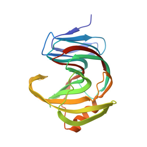
Characterization and structural analysis of a thermophilic GH11 xylanase from compost metatranscriptome.

Yi, Y.Xu, S.Kovalevsky, A.Zhang, X.Liu, D.Wan, Q.

(2021) Appl Microbiol Biotechnol 105: 7757-7767

  • DOI: https://doi.org/10.1007/s00253-021-11587-2
  • Primary Citation Related Structures: 
    7EO6

  • PubMed Abstract: 

    Xylanase is efficient for xylan degradation and widely applied in industries. We found a GH11 family xylanase (Xyn11A) with high thermostability and catalytic activity from compost metatranscriptome. This xylanase has the optimal reaction temperature at 80 °C with the activity of 2907.3 U/mg. The X-ray crystallographic structure shows a typical "right hand" architecture, which is the characteristics of the GH11 family enzymes. Comparing it with the mesophilic XYN II, a well-studied GH11 xylanase from Trichoderma reesei, Xyn11A is more compact with more H-bonds. Our mutagenic results show that the electrostatic interactions in the thumb and palm region of Xyn11A could result in its high thermostability and activity. Introducing a disulfide bond at the N-terminus further increased its optimal reaction temperature to 90 °C with augmented activity. KEY POINTS: • A hyperthermophilic xylanase with high activity was discovered using the metatranscriptomic method. • The mechanisms of thermophilicity and high activity were revealed using X-ray crystallography, mutagenesis, and molecular dynamics simulations. • The thermostability and activity were further improved by introducing a disulfide bond.


  • Organizational Affiliation
    • College of Science, Nanjing Agricultural University, Nanjing, 210095, People's Republic of China.

Macromolecule Content 

  • Total Structure Weight: 43.29 kDa 
  • Atom Count: 3,216 
  • Modeled Residue Count: 381 
  • Deposited Residue Count: 390 
  • Unique protein chains: 1

Macromolecules

Find similar proteins by:|  3D Structure
Entity ID: 1
MoleculeChains  Sequence LengthOrganismDetailsImage
Endo-1,4-beta-xylanase
A, B
195Actinomycetes bacteriumMutation(s): 0 
Gene Names: DIU60_09020
EC: 3.2.1.8

Experimental Data & Validation

Experimental Data

  • Method: X-RAY DIFFRACTION
  • Resolution: 1.90 Å
  • R-Value Free:  0.216 (Depositor), 0.230 (DCC) 
  • R-Value Work:  0.192 (Depositor) 
  • R-Value Observed: 0.193 (Depositor) 
Space Group: P 1
Unit Cell:
Length ( Å )Angle ( ˚ )
a = 35.8α = 74.32
b = 42.476β = 87.4
c = 59.734γ = 86.11
Software Package:
Software NamePurpose
PHENIXrefinement
Aimlessdata scaling
PDB_EXTRACTdata extraction
iMOSFLMdata reduction
MOLREPphasing

Structure Validation

View Full Validation Report


Entry History 

& Funding Information

Deposition Data


Funding OrganizationLocationGrant Number
National Natural Science Foundation of China (NSFC)--

Revision History  (Full details and data files)

  • Version 1.0: 2021-10-13
    Type: Initial release
  • Version 1.1: 2022-02-16
    Changes: Database references
  • Version 1.2: 2023-11-29
    Changes: Data collection, Refinement description