


Identification of Small Molecule Inhibitors of RNase L by Fragment-Based Drug Discovery
Tang, J., Dong, B., Liu, M., Liu, S., Niu, X., Gaughan, C., Asthana, A., Zhou, H., Xu, Z., Zhang, G., Silverman, R.H., Huang, H.(2022) J Med Chem 65: 1445-1457
Experimental Data Snapshot
Starting Model: experimental
View more details
(2022) J Med Chem 65: 1445-1457
Entity ID: 1 | |||||
|---|---|---|---|---|---|
| Molecule | Chains | Sequence Length | Organism | Details | Image |
| Ribonuclease L | A [auth a], B [auth b] | 717 | Sus scrofa | Mutation(s): 0 Gene Names: RNASEL | 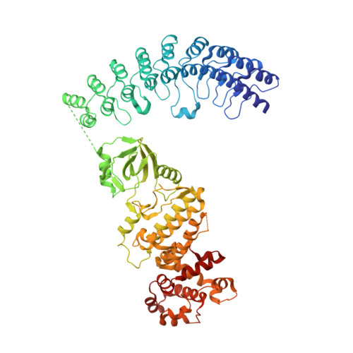 |
UniProt | |||||
Entity Groups | |||||
| Sequence Clusters | 30% Identity50% Identity70% Identity90% Identity95% Identity100% Identity | ||||
| UniProt Group | A5H025 | ||||
Sequence AnnotationsExpand | |||||
Reference Sequence | |||||
| Ligands 3 Unique | |||||
|---|---|---|---|---|---|
| ID | Chains | Name / Formula / InChI Key | 2D Diagram | 3D Interactions | |
| 25L (Subject of Investigation/LOI) Download:Ideal Coordinates CCD File | C [auth a], F [auth b] | [[(2R,3R,4R,5R)-5-(6-aminopurin-9-yl)-4-[[(2R,3R,4R,5R)-5-(6-aminopurin-9-yl)-4-[[(2R,3S,4R,5R)-5-(6-aminopurin-9-yl)-3,4-dihydroxy-oxolan-2-yl]methoxy-hydroxy-phosphoryl]oxy-3-hydroxy-oxolan-2-yl]methoxy-hydroxy-phosphoryl]oxy-3-hydroxy-oxolan-2-yl]methoxy-hydroxy-phosphoryl] phosphono hydrogen phosphate C30 H40 N15 O25 P5 RTAGLZBJCCVJET-UQTMIEBXSA-N |  | ||
| MYC (Subject of Investigation/LOI) Download:Ideal Coordinates CCD File | D [auth a], G [auth b] | 3,5,7-TRIHYDROXY-2-(3,4,5-TRIHYDROXYPHENYL)-4H-CHROMEN-4-ONE C15 H10 O8 IKMDFBPHZNJCSN-UHFFFAOYSA-N |  | ||
| PO4 Download:Ideal Coordinates CCD File | E [auth a] | PHOSPHATE ION O4 P NBIIXXVUZAFLBC-UHFFFAOYSA-K |  | ||
| Length ( Å ) | Angle ( ˚ ) |
|---|---|
| a = 59.608 | α = 90 |
| b = 111.925 | β = 90 |
| c = 264.929 | γ = 90 |
| Software Name | Purpose |
|---|---|
| Aimless | data scaling |
| PHENIX | refinement |
| PDB_EXTRACT | data extraction |
| XDS | data reduction |
| PHENIX | phasing |
| Funding Organization | Location | Grant Number |
|---|---|---|
| National Natural Science Foundation of China (NSFC) | China | 21778808 |


