


Structural and biochemical investigation of UTP cyclohydrolase
Zhang, H., Wei, Y., Lin, L., Liu, J., Chu, R., Cao, P., Ang, E.L., Zhao, H., Yuchi, Z., Zhang, Y.(2021) ACS Catal : 8895-8901
Experimental Data Snapshot
Entity ID: 1 | |||||
|---|---|---|---|---|---|
| Molecule | Chains | Sequence Length | Organism | Details | Image |
| GTP cyclohydrolase II | 425 | Rhodococcus wratislaviensis | Mutation(s): 0 Gene Names: C8E05_0284 | 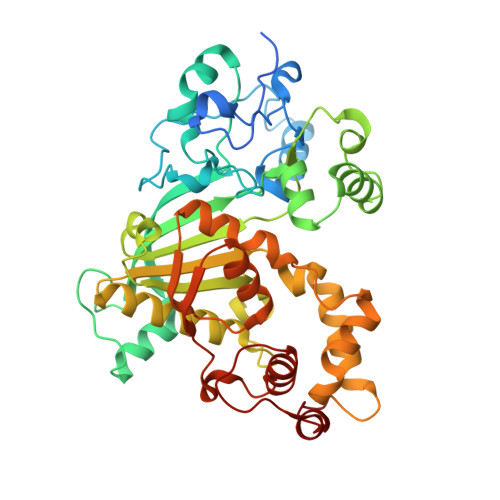 | |
Entity Groups | |||||
| Sequence Clusters | 30% Identity50% Identity70% Identity90% Identity95% Identity100% Identity | ||||
Sequence AnnotationsExpand | |||||
Reference Sequence | |||||
| Ligands 2 Unique | |||||
|---|---|---|---|---|---|
| ID | Chains | Name / Formula / InChI Key | 2D Diagram | 3D Interactions | |
| DUT (Subject of Investigation/LOI) Download:Ideal Coordinates CCD File | C [auth A], E [auth B] | DEOXYURIDINE-5'-TRIPHOSPHATE C9 H15 N2 O14 P3 AHCYMLUZIRLXAA-SHYZEUOFSA-N |  | ||
| ZN (Subject of Investigation/LOI) Download:Ideal Coordinates CCD File | D [auth A], F [auth B] | ZINC ION Zn PTFCDOFLOPIGGS-UHFFFAOYSA-N |  | ||
| Length ( Å ) | Angle ( ˚ ) |
|---|---|
| a = 114.646 | α = 90 |
| b = 114.646 | β = 90 |
| c = 282.713 | γ = 120 |
| Software Name | Purpose |
|---|---|
| PHENIX | refinement |
| HKL-2000 | data scaling |
| PDB_EXTRACT | data extraction |
| HKL-2000 | data reduction |
| PHENIX | phasing |
| Funding Organization | Location | Grant Number |
|---|---|---|
| National Science Foundation (NSF, China) | China | 31870049 |


