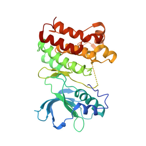
Crystal structure of clinically reported mutations Gly656Arg, Gly656Glu and Asp751His identified in the kinase domain of EphA7.
Chakraborty, S., Varma, A.K.(2021) Biochem Biophys Res Commun 568: 62-67
- PubMed: 34186436 Search on PubMed
- DOI: https://doi.org/10.1016/j.bbrc.2021.06.048
- Primary Citation Related Structures:
7EEC, 7EED, 7EEF - PubMed Abstract:
Erythropoietin producing hepatocellular (Eph) forms the largest family of receptor tyrosine kinases (RTK). As a family, Eph regulates physiological events such as cell-cell interaction, cell migration, and adhesion. The Kinase domain is the catalytic core of the Eph receptor and is highly conserved sequentially. EphA7 has been recently regarded as a cancer driver gene and comprises several clinically important mutations. Three of the EphA7 mutations Gly656Glu, Gly656Arg, and Asp751His, present in the kinase domain, are predicted to be highly pathogenic. Furthermore, Gly656Glu and Gly656Arg are reported to be hotspot mutations. Considering the importance of mutations, crystals structure of EphA7 Gly656Glu, Gly656Arg, and Asp751His mutants has been explored. Changes in folding pattern and intramolecular interactions were observed in mutant structures. Secondary structural changes were observed in the hinge region of EphA7 Gly656Arg and Asp751His structure, affecting the transition of kinase domain between open and closed conformations. EphA7 Asp751His mutant structure shows a distorted nucleotide-binding groove. Differences were observed in hydrogen bonding and hydrophobic interactions between the catalytic and highly conserved DFG motif in the EphA7 mutants, which may influence the catalytic activity of kinase domain.
- Advanced Centre for Treatment, Research and Education in Cancer, Kharghar, Navi Mumbai, Maharashtra, 410210, India; Homi Bhabha National Institute, Training School Complex, Anushaktinagar, Mumbai, Maharashtra, 400094, India.
Organizational Affiliation: