7E6G

Crystal structure of diguanylate cyclase SiaD in complex with its activator SiaC from Pseudomonas aeruginosa


Experimental Data Snapshot

  • Method: X-RAY DIFFRACTION
  • Resolution: 2.65 Å
  • R-Value Free: 0.265 
  • R-Value Work: 0.218 
  • R-Value Observed: 0.220 

wwPDB Validation   3D Report Full Report


Ligand Structure Quality Assessment 


This is version 1.1 of the entry. See complete history


Literature

Structural basis for diguanylate cyclase activation by its binding partner in Pseudomonas aeruginosa .

Chen, G.Zhou, J.Zuo, Y.Huo, W.Peng, J.Li, M.Zhang, Y.Wang, T.Zhang, L.Zhang, L.Liang, H.

(2021) Elife 10

  • DOI: https://doi.org/10.7554/eLife.67289
  • Primary Citation of Related Structures:  
    7E6G

  • PubMed Abstract: 

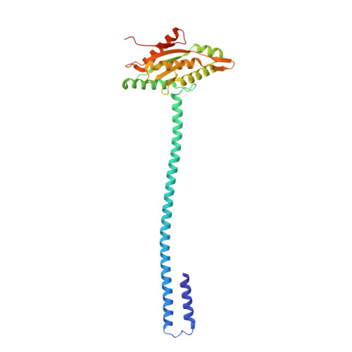
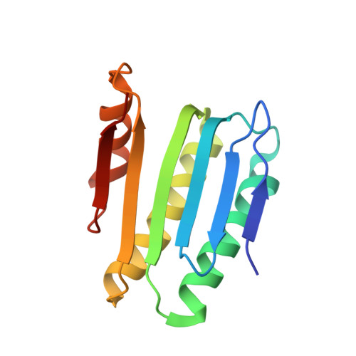
    Cyclic-di-guanosine monophosphate (c-di-GMP) is an important effector associated with acute-chronic infection transition in Pseudomonas aeruginosa . Previously, we reported a signaling network SiaABCD, which regulates biofilm formation by modulating c-di-GMP level. However, the mechanism for SiaD activation by SiaC remains elusive. Here we determine the crystal structure of SiaC-SiaD-GpCpp complex and revealed a unique mirror symmetric conformation: two SiaD form a dimer with long stalk domains, while four SiaC bind to the conserved motifs on the stalks of SiaD and stabilize the conformation for further enzymatic catalysis. Furthermore, SiaD alone exhibits an inactive pentamer conformation in solution, demonstrating that SiaC activates SiaD through a dynamic mechanism of promoting the formation of active SiaD dimers. Mutagenesis assay confirmed that the stalks of SiaD are necessary for its activation. Together, we reveal a novel mechanism for DGC activation, which clarifies the regulatory networks of c-di-GMP signaling.


  • Organizational Affiliation

    Key Laboratory of Resources Biology and Biotechnology in Western China, Ministry of Education, College of Life Sciences, Northwest University, ShaanXi, China.


Macromolecules
Find similar proteins by:  (by identity cutoff)  |  3D Structure
Entity ID: 1
MoleculeChains Sequence LengthOrganismDetailsImage
Putative GGDEF domain protein
A, B
276Pseudomonas aeruginosaMutation(s): 0 
Gene Names: pleD_3pleD_1pleD_4NCTC12951_03415NCTC13437_04900NCTC13621_04785PAMH19_0165RW109_RW109_00771
UniProt
Find proteins for A0A069QEY1 (Pseudomonas aeruginosa)
Explore A0A069QEY1 
Go to UniProtKB:  A0A069QEY1
Entity Groups  
Sequence Clusters30% Identity50% Identity70% Identity90% Identity95% Identity100% Identity
UniProt GroupA0A069QEY1
Sequence Annotations
Expand
  • Reference Sequence
Find similar proteins by:  (by identity cutoff)  |  3D Structure
Entity ID: 2
MoleculeChains Sequence LengthOrganismDetailsImage
DUF1987 domain-containing protein
C, D, E, F
127Pseudomonas aeruginosaMutation(s): 0 
UniProt
Find proteins for A0A072ZHB4 (Pseudomonas aeruginosa)
Explore A0A072ZHB4 
Go to UniProtKB:  A0A072ZHB4
Entity Groups  
Sequence Clusters30% Identity50% Identity70% Identity90% Identity95% Identity100% Identity
UniProt GroupA0A072ZHB4
Sequence Annotations
Expand
  • Reference Sequence
Small Molecules
Experimental Data & Validation

Experimental Data

  • Method: X-RAY DIFFRACTION
  • Resolution: 2.65 Å
  • R-Value Free: 0.265 
  • R-Value Work: 0.218 
  • R-Value Observed: 0.220 
  • Space Group: C 2 2 21
Unit Cell:
Length ( Å )Angle ( ˚ )
a = 82.525α = 90
b = 236.728β = 90
c = 148.627γ = 90
Software Package:
Software NamePurpose
DENZOdata reduction
PHENIXrefinement
PDB_EXTRACTdata extraction
HKL-3000data scaling
PHASERphasing

Structure Validation

View Full Validation Report


Ligand Structure Quality Assessment 


Entry History & Funding Information

Deposition Data


Funding OrganizationLocationGrant Number
National Natural Science Foundation of China (NSFC)China21722802
National Natural Science Foundation of China (NSFC)China91853118

Revision History  (Full details and data files)

  • Version 1.0: 2021-09-22
    Type: Initial release
  • Version 1.1: 2023-11-29
    Changes: Data collection, Refinement description