7E2G

Cryo-EM structure of hDisp1NNN-3C


Experimental Data Snapshot

  • Method: ELECTRON MICROSCOPY
  • Resolution: 3.61 Å
  • Aggregation State: PARTICLE 
  • Reconstruction Method: SINGLE PARTICLE 

wwPDB Validation   3D Report Full Report


This is version 1.0 of the entry. See complete history


Literature

Structural insights into proteolytic activation of the human Dispatched1 transporter for Hedgehog morphogen release.

Li, W.Wang, L.Wierbowski, B.M.Lu, M.Dong, F.Liu, W.Li, S.Wang, P.Salic, A.Gong, X.

(2021) Nat Commun 12: 6966-6966

  • DOI: https://doi.org/10.1038/s41467-021-27257-w
  • Primary Citation of Related Structures:  
    7E2G, 7E2H, 7E2I

  • PubMed Abstract: 

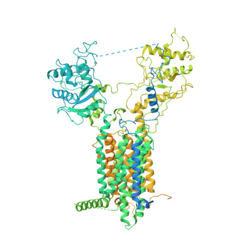
    The membrane protein Dispatched (Disp), which belongs to the RND family of small molecule transporters, is essential for Hedgehog (Hh) signaling, by catalyzing the extracellular release of palmitate- and cholesterol-modified Hh ligands from producing cells. Disp function requires Furin-mediated proteolytic cleavage of its extracellular domain, but how this activates Disp remains obscure. Here, we employ cryo-electron microscopy to determine atomic structures of human Disp1 (hDisp1), before and after cleavage, and in complex with lipid-modified Sonic hedgehog (Shh) ligand. These structures, together with biochemical data, reveal that proteolytic cleavage opens the extracellular domain of hDisp1, removing steric hindrance to Shh binding. Structure-guided functional experiments demonstrate the role of hDisp1-Shh interactions in ligand release. Our results clarify the mechanisms of hDisp1 activation and Shh morphogen release, and highlight how a unique proteolytic cleavage event enabled acquisition of a protein substrate by a member of a family of small molecule transporters.


  • Organizational Affiliation

    Department of Biology, School of Life Sciences, Southern University of Science and Technology, 518055, Shenzhen, Guangdong, China.


Macromolecules
Find similar proteins by:  (by identity cutoff)  |  3D Structure
Entity ID: 1
MoleculeChains Sequence LengthOrganismDetailsImage
Protein dispatched homolog 1,Protein dispatched homolog 1A [auth D]1,518Homo sapiensMutation(s): 3 
Gene Names: DISP1DISPA
Membrane Entity: Yes 
UniProt & NIH Common Fund Data Resources
Find proteins for Q96F81 (Homo sapiens)
Explore Q96F81 
Go to UniProtKB:  Q96F81
PHAROS:  Q96F81
GTEx:  ENSG00000154309 
Entity Groups  
Sequence Clusters30% Identity50% Identity70% Identity90% Identity95% Identity100% Identity
UniProt GroupQ96F81
Sequence Annotations
Expand
  • Reference Sequence
Small Molecules
Ligands 2 Unique
IDChains Name / Formula / InChI Key2D Diagram3D Interactions
Y01 (Subject of Investigation/LOI)
Query on Y01

Download Ideal Coordinates CCD File 
B [auth D]
C [auth D]
D
E [auth D]
F [auth D]
B [auth D],
C [auth D],
D,
E [auth D],
F [auth D],
G [auth D],
H [auth D],
I [auth D],
J [auth D],
K [auth D],
L [auth D]
CHOLESTEROL HEMISUCCINATE
C31 H50 O4
WLNARFZDISHUGS-MIXBDBMTSA-N
NAG (Subject of Investigation/LOI)
Query on NAG

Download Ideal Coordinates CCD File 
M [auth D],
N [auth D],
O [auth D],
P [auth D]
2-acetamido-2-deoxy-beta-D-glucopyranose
C8 H15 N O6
OVRNDRQMDRJTHS-FMDGEEDCSA-N
Experimental Data & Validation

Experimental Data

  • Method: ELECTRON MICROSCOPY
  • Resolution: 3.61 Å
  • Aggregation State: PARTICLE 
  • Reconstruction Method: SINGLE PARTICLE 

Structure Validation

View Full Validation Report


Entry History & Funding Information

Deposition Data


Funding OrganizationLocationGrant Number
Other governmentChina--

Revision History  (Full details and data files)

  • Version 1.0: 2021-12-08
    Type: Initial release