


Crystal structure of uPA in complex with cleaved camostat
Jiang, L.G., Huang, M.D.To be published.
Experimental Data Snapshot
Starting Model: experimental
View more details
Entity ID: 1 | |||||
|---|---|---|---|---|---|
| Molecule | Chains | Sequence Length | Organism | Details | Image |
| Urokinase-type plasminogen activator | A [auth U] | 245 | Homo sapiens | Mutation(s): 2 Gene Names: PLAU EC: 3.4.21.73 | 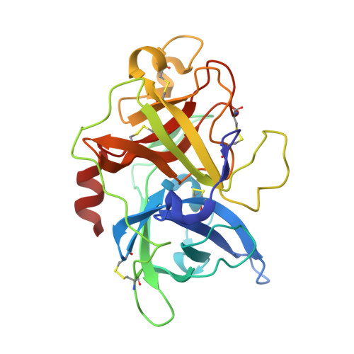 |
UniProt & NIH Common Fund Data Resources | |||||
PHAROS: P00749 GTEx: ENSG00000122861 | |||||
Entity Groups | |||||
| Sequence Clusters | 30% Identity50% Identity70% Identity90% Identity95% Identity100% Identity | ||||
| UniProt Group | P00749 | ||||
Sequence AnnotationsExpand | |||||
Reference Sequence | |||||
| Ligands 2 Unique | |||||
|---|---|---|---|---|---|
| ID | Chains | Name / Formula / InChI Key | 2D Diagram | 3D Interactions | |
| GBS (Subject of Investigation/LOI) Download:Ideal Coordinates CCD File | B [auth U] | 4-carbamimidamidobenzoic acid C8 H9 N3 O2 SXTSBZBQQRIYCU-UHFFFAOYSA-N |  | ||
| PGE Download:Ideal Coordinates CCD File | C [auth U], D [auth U] | TRIETHYLENE GLYCOL C6 H14 O4 ZIBGPFATKBEMQZ-UHFFFAOYSA-N |  | ||
| Length ( Å ) | Angle ( ˚ ) |
|---|---|
| a = 120.317 | α = 90 |
| b = 120.317 | β = 90 |
| c = 42.451 | γ = 120 |
| Software Name | Purpose |
|---|---|
| REFMAC | refinement |
| PDB_EXTRACT | data extraction |
| APEX | data reduction |
| APEX | data scaling |
| MOLREP | phasing |


