


Crystal structure of Catabolite repressor acivator from E. coli in complex with HEPES
Neetu, N., Katiki, M., Kumar, P.To be published.
Experimental Data Snapshot
Starting Model: experimental
View more details
Entity ID: 1 | |||||
|---|---|---|---|---|---|
| Molecule | Chains | Sequence Length | Organism | Details | Image |
| Catabolite repressor/activator | 357 | Escherichia coli 536 | Mutation(s): 0 Gene Names: ECP_0082 | 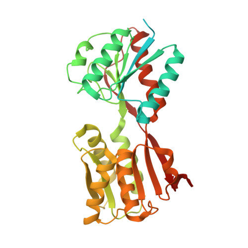 | |
UniProt | |||||
Find proteins for A0A454A0X5 (Escherichia coli O6:K15:H31 (strain 536 / UPEC)) Explore A0A454A0X5 Go to UniProtKB: A0A454A0X5 | |||||
Entity Groups | |||||
| Sequence Clusters | 30% Identity50% Identity70% Identity90% Identity95% Identity100% Identity | ||||
| UniProt Group | A0A454A0X5 | ||||
Sequence AnnotationsExpand | |||||
Reference Sequence | |||||
| Ligands 2 Unique | |||||
|---|---|---|---|---|---|
| ID | Chains | Name / Formula / InChI Key | 2D Diagram | 3D Interactions | |
| EPE (Subject of Investigation/LOI) Download:Ideal Coordinates CCD File | C [auth A], D [auth B], E [auth B] | 4-(2-HYDROXYETHYL)-1-PIPERAZINE ETHANESULFONIC ACID C8 H18 N2 O4 S JKMHFZQWWAIEOD-UHFFFAOYSA-N |  | ||
| GOL Download:Ideal Coordinates CCD File | F [auth B] | GLYCEROL C3 H8 O3 PEDCQBHIVMGVHV-UHFFFAOYSA-N |  | ||
| Length ( Å ) | Angle ( ˚ ) |
|---|---|
| a = 123.932 | α = 90 |
| b = 43.341 | β = 90 |
| c = 108.908 | γ = 90 |
| Software Name | Purpose |
|---|---|
| REFMAC | refinement |
| PDB_EXTRACT | data extraction |
| SCALEPACK | data scaling |
| MOLREP | phasing |
| XDS | data reduction |


