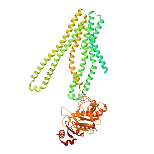
7DNY

Cryo-EM structure of the human ABCB6 (coproporphyrin III-bound)


Experimental Data Snapshot

  • Method: ELECTRON MICROSCOPY
  • Resolution: 3.40 Å
  • Aggregation State: PARTICLE 
  • Reconstruction Method: SINGLE PARTICLE 

wwPDB Validation   3D Report Full Report


This is version 1.2 of the entry. See complete history


Literature

Structural Insights into Porphyrin Recognition by the Human ATP-Binding Cassette Transporter ABCB6.

Kim, S.Lee, S.S.Park, J.G.Kim, J.W.Ju, S.Choi, S.H.Kim, S.Kim, N.J.Hong, S.Kang, J.Y.Jin, M.S.

(2022) Mol Cells 45: 575-587

  • DOI: https://doi.org/10.14348/molcells.2022.0040
  • Primary Citation of Related Structures:  
    7DNY, 7DNZ

  • PubMed Abstract: 

    Human ABCB6 is an ATP-binding cassette transporter that regulates heme biosynthesis by translocating various porphyrins from the cytoplasm into the mitochondria. Here we report the cryo-electron microscopy (cryo-EM) structures of human ABCB6 with its substrates, coproporphyrin III (CPIII) and hemin, at 3.5 and 3.7 Å resolution, respectively. Metalfree porphyrin CPIII binds to ABCB6 within the central cavity, where its propionic acids form hydrogen bonds with the highly conserved Y550. The resulting structure has an overall fold similar to the inward-facing apo structure, but the two nucleotide-binding domains (NBDs) are slightly closer to each other. In contrast, when ABCB6 binds a metal-centered porphyrin hemin in complex with two glutathione molecules (1 hemin: 2 glutathione), the two NBDs end up much closer together, aligning them to bind and hydrolyze ATP more efficiently. In our structures, a glycine-rich and highly flexible "bulge" loop on TM helix 7 undergoes significant conformational changes associated with substrate binding. Our findings suggest that ABCB6 utilizes at least two distinct mechanisms to fine-tune substrate specificity and transport efficiency.


  • Organizational Affiliation

    School of Life Sciences, Gwangju Institute of Science and Technology (GIST), Gwangju 61005, Korea.


Macromolecules
Find similar proteins by:  (by identity cutoff)  |  3D Structure
Entity ID: 1
MoleculeChains Sequence LengthOrganismDetailsImage
ATP-binding cassette sub-family B member 6, mitochondrial
A, B
842Homo sapiensMutation(s): 0 
Gene Names: ABCB6MTABC3PRPUMAT
EC: 7.6.2.5
Membrane Entity: Yes 
UniProt & NIH Common Fund Data Resources
Find proteins for Q9NP58 (Homo sapiens)
Explore Q9NP58 
Go to UniProtKB:  Q9NP58
PHAROS:  Q9NP58
GTEx:  ENSG00000115657 
Entity Groups  
Sequence Clusters30% Identity50% Identity70% Identity90% Identity95% Identity100% Identity
UniProt GroupQ9NP58
Sequence Annotations
Expand
  • Reference Sequence
Experimental Data & Validation

Experimental Data

  • Method: ELECTRON MICROSCOPY
  • Resolution: 3.40 Å
  • Aggregation State: PARTICLE 
  • Reconstruction Method: SINGLE PARTICLE 

Structure Validation

View Full Validation Report


Entry History & Funding Information

Deposition Data


Funding OrganizationLocationGrant Number
National Research Foundation (NRF, Korea)Korea, Republic OfNRF-2017M3A9F6029753
National Research Foundation (NRF, Korea)Korea, Republic OfNRF-2020R1I1A1A01072077
National Research Foundation (NRF, Korea)Korea, Republic OfNRF-2019M3E5D6066058
National Research Foundation (NRF, Korea)Korea, Republic OfNRF-2019R1A6A1A10073887

Revision History  (Full details and data files)

  • Version 1.0: 2022-06-22
    Type: Initial release
  • Version 1.1: 2022-08-31
    Changes: Database references
  • Version 1.2: 2024-06-05
    Changes: Data collection