7DN8

Crystal structure of Salmonella effector SopF in complex with ARF1


Experimental Data Snapshot

  • Method: X-RAY DIFFRACTION
  • Resolution: 2.61 Å
  • R-Value Free: 0.240 
  • R-Value Work: 0.201 
  • R-Value Observed: 0.202 

wwPDB Validation   3D Report Full Report


Ligand Structure Quality Assessment 


This is version 1.1 of the entry. See complete history


Literature

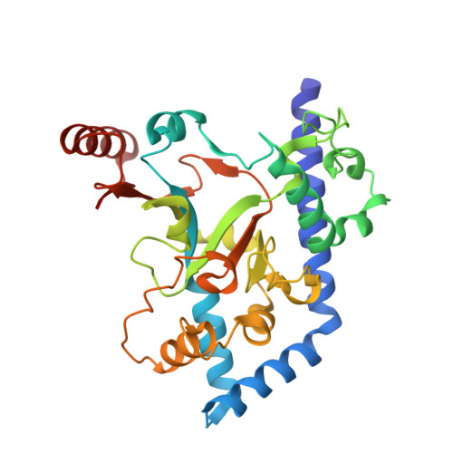
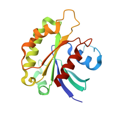
ARF GTPases activate Salmonella effector SopF to ADP-ribosylate host V-ATPase and inhibit endomembrane damage-induced autophagy.

Xu, Y.Cheng, S.Zeng, H.Zhou, P.Ma, Y.Li, L.Liu, X.Shao, F.Ding, J.

(2022) Nat Struct Mol Biol 29: 67-77

  • DOI: https://doi.org/10.1038/s41594-021-00710-6
  • Primary Citation of Related Structures:  
    7DN8, 7DN9

  • PubMed Abstract: 

    Selective autophagy helps eukaryotes to cope with endogenous dangers or foreign invaders; its initiation often involves membrane damage. By studying a Salmonella effector SopF, we recently identified the vacuolar ATPase (V-ATPase)-ATG16L1 axis that initiates bacteria-induced autophagy. Here we show that SopF is an ADP-ribosyltransferase specifically modifying Gln124 of ATP6V0C in V-ATPase. We identify GTP-bound ADP-ribosylation factor (ARF) GTPases as a cofactor required for SopF functioning. Crystal structures of SopF-ARF1 complexes not only reveal structural basis of SopF ADP-ribosyltransferase activity but also a unique effector-binding mode adopted by ARF GTPases. Further, the N terminus of ARF1, although dispensable for high-affinity binding to SopF, is critical for activating SopF to modify ATP6V0C. Moreover, lysosome or Golgi damage-induced autophagic LC3 activation is inhibited by SopF or Q124A mutation of ATP6V0C, thus also mediated by the V-ATPase-ATG16L1 axis. In this process, the V-ATPase functions to sense membrane damages, which can be uncoupled from its proton-pumping activity.


  • Organizational Affiliation

    National Institute of Biological Sciences, Beijing, China.


Macromolecules
Find similar proteins by:  (by identity cutoff)  |  3D Structure
Entity ID: 1
MoleculeChains Sequence LengthOrganismDetailsImage
Putative cytoplasmic proteinA [auth C],
C [auth A],
E,
G
305Salmonella enterica subsp. enterica serovar Typhimurium str. LT2Mutation(s): 0 
Gene Names: STM1239
UniProt
Find proteins for Q8ZPY9 (Salmonella typhimurium (strain LT2 / SGSC1412 / ATCC 700720))
Explore Q8ZPY9 
Go to UniProtKB:  Q8ZPY9
Entity Groups  
Sequence Clusters30% Identity50% Identity70% Identity90% Identity95% Identity100% Identity
UniProt GroupQ8ZPY9
Sequence Annotations
Expand
  • Reference Sequence
Find similar proteins by:  (by identity cutoff)  |  3D Structure
Entity ID: 2
MoleculeChains Sequence LengthOrganismDetailsImage
ADP-ribosylation factor 1B [auth D],
D [auth B],
F,
H
169Homo sapiensMutation(s): 1 
Gene Names: ARF1
UniProt & NIH Common Fund Data Resources
Find proteins for P84077 (Homo sapiens)
Explore P84077 
Go to UniProtKB:  P84077
PHAROS:  P84077
GTEx:  ENSG00000143761 
Entity Groups  
Sequence Clusters30% Identity50% Identity70% Identity90% Identity95% Identity100% Identity
UniProt GroupP84077
Sequence Annotations
Expand
  • Reference Sequence
Experimental Data & Validation

Experimental Data

  • Method: X-RAY DIFFRACTION
  • Resolution: 2.61 Å
  • R-Value Free: 0.240 
  • R-Value Work: 0.201 
  • R-Value Observed: 0.202 
  • Space Group: P 1 21 1
Unit Cell:
Length ( Å )Angle ( ˚ )
a = 104.737α = 90
b = 104.902β = 104.88
c = 105.603γ = 90
Software Package:
Software NamePurpose
PHENIXrefinement
XDSdata reduction
XSCALEdata scaling
PHENIXphasing

Structure Validation

View Full Validation Report


Ligand Structure Quality Assessment 


Entry History 

Deposition Data

Revision History  (Full details and data files)

  • Version 1.0: 2021-12-15
    Type: Initial release
  • Version 1.1: 2022-12-28
    Changes: Database references