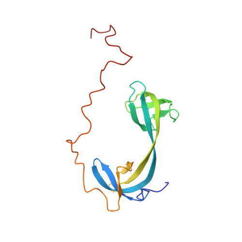
Structural basis for the DNA-binding activity of human ARID4B Tudor domain.
Ren, J., Yao, H., Hu, W., Perrett, S., Gong, W., Feng, Y.(2021) J Biological Chem 296: 100506-100506
- PubMed: 33675746 Search on PubMedSearch on PubMed Central
- DOI: https://doi.org/10.1016/j.jbc.2021.100506
- Primary Citation Related Structures:
7DM4 - PubMed Abstract:
Human ARID4A and ARID4B are homologous proteins that are important in controlling gene expression and epigenetic regulation but have distinct functions. Previous studies have shown that the N-terminal domain of ARID4A is an unusual interdigitated double Tudor domain with DNA-binding activity. However, how the Tudor domain of ARID4B differs from that of ARID4A remains unknown. Here, we found that the ARID4B Tudor domain has significantly weaker DNA affinity than the ARID4A Tudor domain despite sharing more than 80% sequence identity. Structure determination and DNA titration analysis indicated that the ARID4B Tudor domain is also an interdigitated double Tudor domain with a DNA-binding surface similar to ARID4A. We identified a residue close to the DNA-binding site of the Tudor domain that differs between ARID4A and ARID4B. The Leu50 in ARID4A is Glu50 in ARID4B, and the latter forms salt bridges with two lysine residues at the DNA-binding surface. This causes a decrease in the strength of positive charge, thus reducing DNA-binding affinity while significantly increasing protein stability. We also found that a C-terminal extension region enhances the DNA-binding affinity of the ARID4B Tudor domain. This C-terminal extension is disordered and contains a positively charged RGR motif, providing an additional DNA-binding site. Finally, sequence and phylogenetic analyses indicated that the residue differences and the presence of the RGR extension region are conserved. These results provide new insight into the functional differences between ARID4A and ARID4B proteins, as well as elucidating the function of the disordered regions in these proteins.
- National Laboratory of Biomacromolecules, CAS Center for Excellence in Biomacromolecules, Institute of Biophysics, Chinese Academy of Sciences, Beijing, China; University of Chinese Academy of Sciences, Beijing, China.
Organizational Affiliation: