7DL7 | pdb_00007dl7

The wild-type structure of 3,5-DAHDHcca


Experimental Data Snapshot

  • Method: X-RAY DIFFRACTION
  • Resolution: 2.30 Å
  • R-Value Free: 
    0.257 (Depositor), 0.261 (DCC) 
  • R-Value Work: 
    0.226 (Depositor), 0.229 (DCC) 
  • R-Value Observed: 
    0.227 (Depositor) 

wwPDB Validation 3D Report Full Report

Validation slider image for 7DL7

Ligand Structure Quality Assessment 


This is version 1.1 of the entry. See complete history

Literature

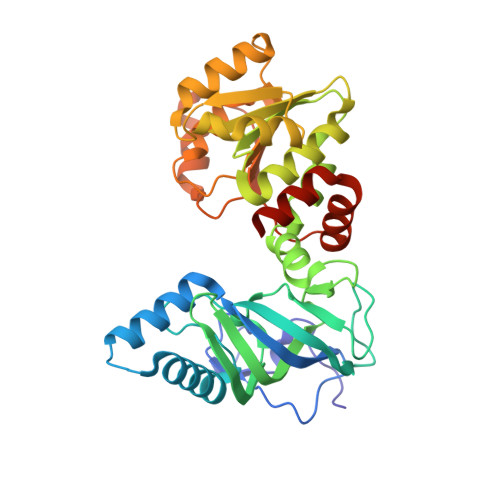
Crystal Structures and Catalytic Mechanism of l-erythro-3,5-Diaminohexanoate Dehydrogenase and Rational Engineering for Asymmetric Synthesis of beta-Amino Acids.

Liu, N.Wu, L.Feng, J.Sheng, X.Li, J.Chen, X.Li, J.Liu, W.Zhou, J.Wu, Q.Zhu, D.

(2021) Angew Chem Int Ed Engl 60: 10203-10210

  • DOI: https://doi.org/10.1002/anie.202017225
  • Primary Citation Related Structures: 
    7DL0, 7DL1, 7DL3, 7DL7

  • PubMed Abstract: 

    Amino acid dehydrogenases (AADHs) have shown considerable potential as biocatalysts in the asymmetric synthesis of chiral amino acids. However, compared to the widely studied α-AADHs, limited knowledge is available about β-AADHs that enable the synthesis of β-amino acids. Herein, we report the crystal structures of a l-erythro-3,5-diaminohexanoate dehydrogenase and its variants, the only known member of β-AADH family. Crystal structure analysis, site-directed mutagenesis studies and quantum chemical calculations revealed the differences in the substrate binding and catalytic mechanism from α-AADHs. A number of rationally engineered variants were then obtained with improved activity (by 110-800 times) toward various aliphatic β-amino acids without an enantioselectivity trade-off. Two β-amino acids were prepared by using the outstanding variants with excellent enantioselectivity (>99 % ee) and high isolated yields (86-87 %). These results provide important insights into the molecular mechanism of 3,5-DAHDH, and establish a solid foundation for further design of β-AADHs for the asymmetric synthesis of β-amino acids.


  • Organizational Affiliation
    • National Engineering Laboratory for Industrial Enzymes and Tianjin Engineering Research Center of Biocatalytic Technology, Tianjin Institute of Industrial Biotechnology, Chinese Academy of Sciences, and, National Technology Innovation Center for Synthetic Biology, Tianjin, 300308, China.

Macromolecule Content 

  • Total Structure Weight: 316.13 kDa 
  • Atom Count: 21,840 
  • Modeled Residue Count: 2,803 
  • Deposited Residue Count: 2,864 
  • Unique protein chains: 1

Macromolecules

Find similar proteins by:|  3D Structure
Entity ID: 1
MoleculeChains  Sequence LengthOrganismDetailsImage
3,5-diaminohexanoate dehydrogenase
A, B, C, D, E
A, B, C, D, E, F, G, H
358Candidatus Cloacimonas acidaminovorans str. EvryMutation(s): 0 
Gene Names: kddCLOAM1348
EC: 1.4.1.11
UniProt
Find proteins for B0VJ11 (Cloacimonas acidaminovorans (strain Evry))
Explore B0VJ11 
Go to UniProtKB:  B0VJ11
Entity Groups
Sequence Clusters30% Identity50% Identity70% Identity90% Identity95% Identity100% Identity
UniProt GroupB0VJ11
Sequence Annotations
Expand
Reference Sequence

Small Molecules

Ligands 7 Unique
IDChains Name / Formula / InChI Key2D Diagram3D Interactions
TRS
(Subject of Investigation/LOI)

Query on TRS


Download:Ideal Coordinates CCD File
KA [auth F]2-AMINO-2-HYDROXYMETHYL-PROPANE-1,3-DIOL
C4 H12 N O3
LENZDBCJOHFCAS-UHFFFAOYSA-O
PEG
(Subject of Investigation/LOI)

Query on PEG


Download:Ideal Coordinates CCD File
I [auth A],
N [auth B],
U [auth C],
UA [auth H]
DI(HYDROXYETHYL)ETHER
C4 H10 O3
MTHSVFCYNBDYFN-UHFFFAOYSA-N
SO4

Query on SO4


Download:Ideal Coordinates CCD File
MA [auth F],
T [auth B]
SULFATE ION
O4 S
QAOWNCQODCNURD-UHFFFAOYSA-L
GOL
(Subject of Investigation/LOI)

Query on GOL


Download:Ideal Coordinates CCD File
DA [auth D]GLYCEROL
C3 H8 O3
PEDCQBHIVMGVHV-UHFFFAOYSA-N
EDO

Query on EDO


Download:Ideal Coordinates CCD File
AA [auth D]
BA [auth D]
FA [auth E]
HA [auth F]
IA [auth F]
AA [auth D],
BA [auth D],
FA [auth E],
HA [auth F],
IA [auth F],
J [auth A],
JA [auth F],
K [auth A],
L [auth A],
NA [auth G],
O [auth B],
OA [auth G],
P [auth B],
PA [auth G],
Q [auth B],
QA [auth G],
R [auth B],
RA [auth G],
S [auth B],
V [auth C],
VA [auth H],
W [auth C],
WA [auth H],
X [auth C],
XA [auth H],
Y [auth C],
YA [auth H]
1,2-ETHANEDIOL
C2 H6 O2
LYCAIKOWRPUZTN-UHFFFAOYSA-N
CA

Query on CA


Download:Ideal Coordinates CCD File
EA [auth D],
TA [auth G]
CALCIUM ION
Ca
BHPQYMZQTOCNFJ-UHFFFAOYSA-N
CL

Query on CL


Download:Ideal Coordinates CCD File
AB [auth H]
CA [auth D]
GA [auth E]
LA [auth F]
M [auth A]
AB [auth H],
CA [auth D],
GA [auth E],
LA [auth F],
M [auth A],
SA [auth G],
Z [auth C],
ZA [auth H]
CHLORIDE ION
Cl
VEXZGXHMUGYJMC-UHFFFAOYSA-M

Experimental Data & Validation

Experimental Data

  • Method: X-RAY DIFFRACTION
  • Resolution: 2.30 Å
  • R-Value Free:  0.257 (Depositor), 0.261 (DCC) 
  • R-Value Work:  0.226 (Depositor), 0.229 (DCC) 
  • R-Value Observed: 0.227 (Depositor) 
Space Group: P 1
Unit Cell:
Length ( Å )Angle ( ˚ )
a = 89.604α = 87.328
b = 89.889β = 70.97
c = 123.693γ = 80.562
Software Package:
Software NamePurpose
PHENIXrefinement
HKL-2000data reduction
HKL-2000data scaling
PHENIXphasing

Structure Validation

View Full Validation Report


Ligand Structure Quality Assessment 


Entry History 

Deposition Data

Revision History  (Full details and data files)

  • Version 1.0: 2021-09-29
    Type: Initial release
  • Version 1.1: 2024-05-29
    Changes: Data collection