7DJR | pdb_00007djr

Crystal structure of SARS-CoV-2 main protease (no ligand)


Experimental Data Snapshot

  • Method: X-RAY DIFFRACTION
  • Resolution: 1.45 Å
  • R-Value Free: 
    0.201 (Depositor), 0.202 (DCC) 
  • R-Value Work: 
    0.166 (Depositor), 0.167 (DCC) 
  • R-Value Observed: 
    0.168 (Depositor) 

Starting Model: experimental
View more details

wwPDB Validation 3D Report Full Report

Validation slider image for 7DJR

This is version 1.3 of the entry. See complete history

Literature

Interaction of 8-anilinonaphthalene-1-sulfonate with SARS-CoV-2 main protease and its application as a fluorescent probe for inhibitor identification.

Deetanya, P.Hengphasatporn, K.Wilasluck, P.Shigeta, Y.Rungrotmongkol, T.Wangkanont, K.

(2021) Comput Struct Biotechnol J 19: 3364-3371

  • DOI: https://doi.org/10.1016/j.csbj.2021.05.053
  • Primary Citation Related Structures: 
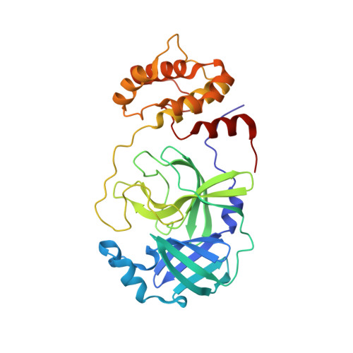
    7DJR

  • PubMed Abstract: 

    The 3C-like main protease of SARS-CoV-2 (3CL Pro ) is responsible for the cleavage of the viral polyprotein. This process is essential for the viral life cycle. Therefore, 3CL Pro is a promising target to develop antiviral drugs for COVID-19 prevention and treatment. Traditional enzymatic assays for the identification of 3CL Pro inhibitors rely on peptide-based colorimetric or fluorogenic substrates. However, the COVID-19 pandemic has limit or delay access to these substrates, especially for researchers in developing countries attempting to screen natural product libraries. We explored the use of the fluorescent probe 8-anilinonaphthalene-1-sulfonate (ANS) as an alternative assay for inhibitor identification. Fluorescence enhancement upon binding of ANS to 3CL Pro was observed, and this interaction was competitive with a peptide substrate. The utility of ANS-based competitive binding assay to identify 3CL Pro inhibitors was demonstrated with the flavonoid natural products baicalein and rutin. The molecular nature of ANS and rutin interaction with 3CL Pro was explored with molecular modeling. Our results suggested that ANS could be employed in a competitive binding assay to facilitate the identification of novel SARS-CoV-2 antiviral compounds.


  • Organizational Affiliation
    • Center of Excellence for Molecular Biology and Genomics of Shrimp, Department of Biochemistry, Faculty of Science, Chulalongkorn University, Bangkok 10330, Thailand.

Macromolecule Content 

  • Total Structure Weight: 34.14 kDa 
  • Atom Count: 2,935 
  • Modeled Residue Count: 305 
  • Deposited Residue Count: 306 
  • Unique protein chains: 1

Macromolecules

Find similar proteins by:|  3D Structure
Entity ID: 1
MoleculeChains  Sequence LengthOrganismDetailsImage
3C-like proteinase306Severe acute respiratory syndrome coronavirus 2Mutation(s): 0 
EC: 3.4.22.69
UniProt
Find proteins for P0DTD1 (Severe acute respiratory syndrome coronavirus 2)
Explore P0DTD1 
Go to UniProtKB:  P0DTD1
Entity Groups
Sequence Clusters30% Identity50% Identity70% Identity90% Identity95% Identity100% Identity
UniProt GroupP0DTD1
Sequence Annotations
Expand
Reference Sequence

Experimental Data & Validation

Experimental Data

  • Method: X-RAY DIFFRACTION
  • Resolution: 1.45 Å
  • R-Value Free:  0.201 (Depositor), 0.202 (DCC) 
  • R-Value Work:  0.166 (Depositor), 0.167 (DCC) 
  • R-Value Observed: 0.168 (Depositor) 
Space Group: C 1 2 1
Unit Cell:
Length ( Å )Angle ( ˚ )
a = 113.225α = 90
b = 53.017β = 103.06
c = 44.692γ = 90
Software Package:
Software NamePurpose
XDSdata reduction
Aimlessdata scaling
PHASERphasing
PHENIXrefinement
PDB_EXTRACTdata extraction

Structure Validation

View Full Validation Report


Entry History 

Deposition Data

Revision History  (Full details and data files)

  • Version 1.0: 2021-06-16
    Type: Initial release
  • Version 1.1: 2021-06-23
    Changes: Database references
  • Version 1.2: 2021-06-30
    Changes: Database references
  • Version 1.3: 2023-11-29
    Changes: Data collection, Database references, Refinement description