7DGD | pdb_00007dgd

apo state of class C GPCR


Experimental Data Snapshot

  • Method: ELECTRON MICROSCOPY
  • Resolution: 3.96 Å
  • Aggregation State: PARTICLE 
  • Reconstruction Method: SINGLE PARTICLE 

wwPDB Validation 3D Report Full Report

Validation slider image for 7DGD

This is version 1.1 of the entry. See complete history

Literature

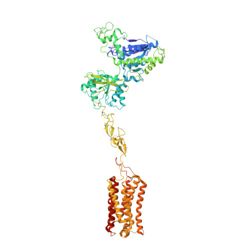
Structural insights into the activation initiation of full-length mGlu1.

Zhang, J.Qu, L.Wu, L.Tang, X.Luo, F.Xu, W.Xu, Y.Liu, Z.J.Hua, T.

(2021) Protein Cell 12: 662-667

  • DOI: https://doi.org/10.1007/s13238-020-00808-5
  • Primary Citation Related Structures: 
    7DGD, 7DGE

  • Organizational Affiliation
    • iHuman Institute, ShanghaiTech University, Shanghai, 201210, China.

Macromolecule Content 

  • Total Structure Weight: 186.6 kDa 
  • Atom Count: 12,381 
  • Modeled Residue Count: 1,572 
  • Deposited Residue Count: 1,666 
  • Unique protein chains: 1

Macromolecules

Find similar proteins by:|  3D Structure
Entity ID: 1
MoleculeChains  Sequence LengthOrganismDetailsImage
Metabotropic glutamate receptor 1
A, B
833Homo sapiensMutation(s): 0 
Gene Names: GRM1GPRC1AMGLUR1
Membrane Entity: Yes 
UniProt & NIH Common Fund Data Resources
Find proteins for Q13255 (Homo sapiens)
Explore Q13255 
Go to UniProtKB:  Q13255
PHAROS:  Q13255
GTEx:  ENSG00000152822 
Entity Groups
Sequence Clusters30% Identity50% Identity70% Identity90% Identity95% Identity100% Identity
UniProt GroupQ13255
Sequence Annotations
Expand
Reference Sequence

Experimental Data & Validation

Experimental Data

  • Method: ELECTRON MICROSCOPY
  • Resolution: 3.96 Å
  • Aggregation State: PARTICLE 
  • Reconstruction Method: SINGLE PARTICLE 

Structure Validation

View Full Validation Report


Entry History 

Deposition Data

Revision History  (Full details and data files)

  • Version 1.0: 2021-09-22
    Type: Initial release
  • Version 1.1: 2024-11-06
    Changes: Data collection, Structure summary