7DDP

Cryo-EM structure of human ACE2 and GX/P2V/2017 RBD


Experimental Data Snapshot

  • Method: ELECTRON MICROSCOPY
  • Resolution: 3.40 Å
  • Aggregation State: PARTICLE 
  • Reconstruction Method: SINGLE PARTICLE 

wwPDB Validation   3D Report Full Report


This is version 1.1 of the entry. See complete history


Literature

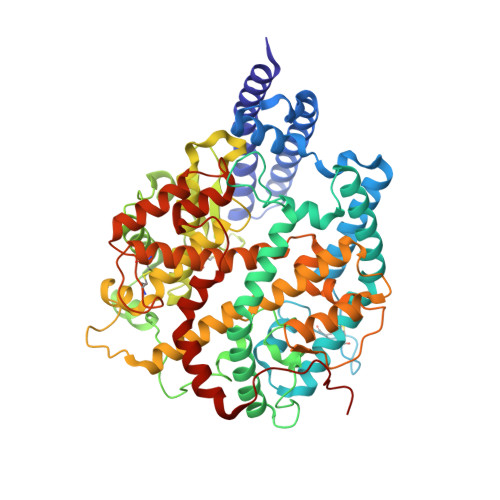
Molecular basis of cross-species ACE2 interactions with SARS-CoV-2-like viruses of pangolin origin.

Niu, S.Wang, J.Bai, B.Wu, L.Zheng, A.Chen, Q.Du, P.Han, P.Zhang, Y.Jia, Y.Qiao, C.Qi, J.Tian, W.X.Wang, H.W.Wang, Q.Gao, G.F.

(2021) EMBO J 40: e107786-e107786

  • DOI: https://doi.org/10.15252/embj.2021107786
  • Primary Citation of Related Structures:  
    7DDP

  • PubMed Abstract: 

    Pangolins have been suggested as potential reservoir of zoonotic viruses, including SARS-CoV-2 causing the global COVID-19 outbreak. Here, we study the binding of two SARS-CoV-2-like viruses isolated from pangolins, GX/P2V/2017 and GD/1/2019, to human angiotensin-converting enzyme 2 (hACE2), the receptor of SARS-CoV-2. We find that the spike protein receptor-binding domain (RBD) of pangolin CoVs binds to hACE2 as efficiently as the SARS-CoV-2 RBD in vitro. Furthermore, incorporation of pangolin CoV RBDs allows entry of pseudotyped VSV particles into hACE2-expressing cells. A screen for binding of pangolin CoV RBDs to ACE2 orthologs from various species suggests a broader host range than that of SARS-CoV-2. Additionally, cryo-EM structures of GX/P2V/2017 and GD/1/2019 RBDs in complex with hACE2 show their molecular binding in modes similar to SARS-CoV-2 RBD. Introducing the Q498H substitution found in pangolin CoVs into the SARS-CoV-2 RBD expands its binding capacity to ACE2 homologs of mouse, rat, and European hedgehog. These findings suggest that these two pangolin CoVs may infect humans, highlighting the necessity of further surveillance of pangolin CoVs.


  • Organizational Affiliation

    College of Veterinary Medicine, Shanxi Agricultural University, Jinzhong, China.


Macromolecules
Find similar proteins by:  (by identity cutoff)  |  3D Structure
Entity ID: 1
MoleculeChains Sequence LengthOrganismDetailsImage
Angiotensin-converting enzyme 2597Homo sapiensMutation(s): 0 
Gene Names: ACE2UNQ868/PRO1885
EC: 3.4.17.23 (PDB Primary Data), 3.4.17 (PDB Primary Data)
UniProt & NIH Common Fund Data Resources
Find proteins for Q9BYF1 (Homo sapiens)
Explore Q9BYF1 
Go to UniProtKB:  Q9BYF1
PHAROS:  Q9BYF1
GTEx:  ENSG00000130234 
Entity Groups  
Sequence Clusters30% Identity50% Identity70% Identity90% Identity95% Identity100% Identity
UniProt GroupQ9BYF1
Sequence Annotations
Expand
  • Reference Sequence
Find similar proteins by:  (by identity cutoff)  |  3D Structure
Entity ID: 2
MoleculeChains Sequence LengthOrganismDetailsImage
Spike protein S1B [auth C]209Pangolin coronavirusMutation(s): 0 
UniProt
Find proteins for A0A6G6A2Q2 (Pangolin coronavirus)
Explore A0A6G6A2Q2 
Go to UniProtKB:  A0A6G6A2Q2
Entity Groups  
Sequence Clusters30% Identity50% Identity70% Identity90% Identity95% Identity100% Identity
UniProt GroupA0A6G6A2Q2
Sequence Annotations
Expand
  • Reference Sequence
Experimental Data & Validation

Experimental Data

  • Method: ELECTRON MICROSCOPY
  • Resolution: 3.40 Å
  • Aggregation State: PARTICLE 
  • Reconstruction Method: SINGLE PARTICLE 

Structure Validation

View Full Validation Report


Entry History 

Deposition Data

Revision History  (Full details and data files)

  • Version 1.0: 2021-05-19
    Type: Initial release
  • Version 1.1: 2022-02-16
    Changes: Database references