7DCV | pdb_00007dcv

Structure of the transmembrane domain of human PD-L1


Experimental Data Snapshot

  • Method: SOLUTION NMR
  • Conformers Calculated: 200 
  • Conformers Submitted: 15 
  • Selection Criteria: structures with the lowest energy 

wwPDB Validation 3D Report Full Report

Validation slider image for 7DCV

This is version 1.1 of the entry. See complete history

Literature

Structure of the transmembrane domain of human PD-L1

Maorong, W.Bo, O.

(2022) Sci Adv 

Macromolecule Content 

  • Total Structure Weight: 6.57 kDa 
  • Atom Count: 463 
  • Modeled Residue Count: 59 
  • Deposited Residue Count: 59 
  • Unique protein chains: 1

Macromolecules

Find similar proteins by:|  3D Structure
Entity ID: 1
MoleculeChains  Sequence LengthOrganismDetailsImage
Programmed cell death 1 ligand 159Homo sapiensMutation(s): 4 
Gene Names: CD274B7H1PDCD1L1PDCD1LG1PDL1
Membrane Entity: Yes 
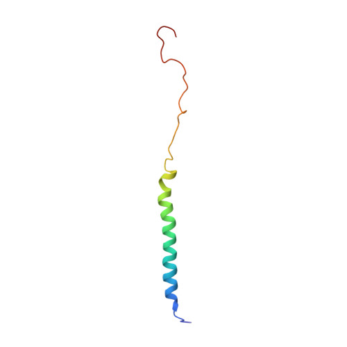
UniProt & NIH Common Fund Data Resources
Find proteins for Q9NZQ7 (Homo sapiens)
Explore Q9NZQ7 
Go to UniProtKB:  Q9NZQ7
PHAROS:  Q9NZQ7
GTEx:  ENSG00000120217 
Entity Groups
Sequence Clusters30% Identity50% Identity70% Identity90% Identity95% Identity100% Identity
UniProt GroupQ9NZQ7
Sequence Annotations
Expand
Reference Sequence

Experimental Data & Validation

Experimental Data

  • Method: SOLUTION NMR
  • Conformers Calculated: 200 
  • Conformers Submitted: 15 
  • Selection Criteria: structures with the lowest energy 

Structure Validation

View Full Validation Report


Entry History 

& Funding Information

Deposition Data


Funding OrganizationLocationGrant Number
National Natural Science Foundation of China (NSFC)China31861133009

Revision History  (Full details and data files)

  • Version 1.0: 2022-08-31
    Type: Initial release
  • Version 1.1: 2024-05-15
    Changes: Data collection, Database references