7DCJ | pdb_00007dcj

Crystal structure of HSF1 DNA-binding domain in complex with 2-site HSE DNA in the head-to-head orientation


Experimental Data Snapshot

  • Method: X-RAY DIFFRACTION
  • Resolution: 2.00 Å
  • R-Value Free: 
    0.222 (Depositor), 0.216 (DCC) 
  • R-Value Work: 
    0.183 (Depositor), 0.180 (DCC) 
  • R-Value Observed: 
    0.185 (Depositor) 

Starting Model: experimental
View more details

wwPDB Validation 3D Report Full Report

Validation slider image for 7DCJ

This is version 1.2 of the entry. See complete history

Literature

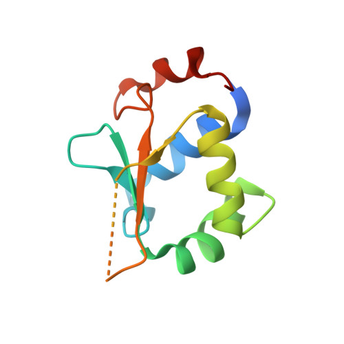
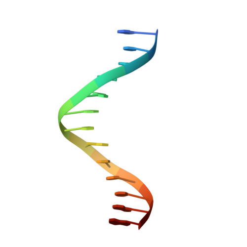
Structures of heat shock factor trimers bound to DNA.

Feng, N.Feng, H.Wang, S.Punekar, A.S.Ladenstein, R.Wang, D.C.Zhang, Q.Ding, J.Liu, W.

(2021) iScience 24: 102951-102951

  • DOI: https://doi.org/10.1016/j.isci.2021.102951
  • Primary Citation Related Structures: 
    7DCJ, 7DCS, 7DCT, 7DCU

  • PubMed Abstract: 

    Heat shock factor 1 (HSF1) and 2 (HSF2) play distinct but overlapping regulatory roles in maintaining cellular proteostasis or mediating cell differentiation and development. Upon activation, both HSFs trimerize and bind to heat shock elements (HSEs) present in the promoter region of target genes. Despite structural insights gained from recent studies, structures reflecting the physiological architecture of this transcriptional machinery remains to be determined. Here, we present co-crystal structures of human HSF1 and HSF2 trimers bound to DNA, which reveal a triangular arrangement of the three DNA-binding domains (DBDs) with protein-protein interactions largely mediated by the wing domain. Two structural properties, different flexibility of the wing domain and local DNA conformational changes induced by HSF binding, seem likely to contribute to the subtle differential specificity between HSF1 and HSF2. Besides, two more structures showing DBDs bound to "two-site" head-to-head HSEs were determined as additions to the published tail-to-tail dimer-binding structures.


  • Organizational Affiliation
    • National Laboratory of Biomacromolecules, Institute of Biophysics, Chinese Academy of Sciences, Beijing 100101, China.

Macromolecule Content 

  • Total Structure Weight: 35.04 kDa 
  • Atom Count: 2,524 
  • Modeled Residue Count: 230 
  • Deposited Residue Count: 254 
  • Unique protein chains: 1
  • Unique nucleic acid chains: 1

Macromolecules


Find similar proteins by:|  3D Structure
Entity ID: 1
MoleculeChains  Sequence LengthOrganismDetailsImage
Heat shock factor protein 1
A, B
113Homo sapiensMutation(s): 0 
Gene Names: HSF1HSTF1
UniProt & NIH Common Fund Data Resources
Find proteins for Q00613 (Homo sapiens)
Explore Q00613 
Go to UniProtKB:  Q00613
PHAROS:  Q00613
GTEx:  ENSG00000185122 
Entity Groups
Sequence Clusters30% Identity50% Identity70% Identity90% Identity95% Identity100% Identity
UniProt GroupQ00613
Sequence Annotations
Expand
Reference Sequence
Find similar nucleic acids by:  Sequence
Entity ID: 2
MoleculeChains LengthOrganismImage
DNA (5'-D(*GP*CP*CP*GP*AP*AP*TP*AP*TP*TP*CP*GP*G)-3')
C, D
14Homo sapiens
Sequence Annotations
Expand
Reference Sequence

Experimental Data & Validation

Experimental Data

  • Method: X-RAY DIFFRACTION
  • Resolution: 2.00 Å
  • R-Value Free:  0.222 (Depositor), 0.216 (DCC) 
  • R-Value Work:  0.183 (Depositor), 0.180 (DCC) 
  • R-Value Observed: 0.185 (Depositor) 
Space Group: P 21 21 21
Unit Cell:
Length ( Å )Angle ( ˚ )
a = 42.221α = 90
b = 62.765β = 90
c = 97.345γ = 90
Software Package:
Software NamePurpose
PHENIXrefinement
PDB_EXTRACTdata extraction
HKL-2000data reduction
HKL-2000data scaling
PHENIXphasing

Structure Validation

View Full Validation Report


Entry History 

Deposition Data

Revision History  (Full details and data files)

  • Version 1.0: 2021-07-14
    Type: Initial release
  • Version 1.1: 2021-09-15
    Changes: Database references, Structure summary
  • Version 1.2: 2023-11-29
    Changes: Data collection, Refinement description