7DB7 | pdb_00007db7

Crystal structure of Mycobacterium tuberculosis phenylalanyl-tRNA synthetase in complex with compound GDI05-001


Experimental Data Snapshot

  • Method: X-RAY DIFFRACTION
  • Resolution: 2.71 Å
  • R-Value Free: 
    0.219 (Depositor), 0.218 (DCC) 
  • R-Value Work: 
    0.193 (Depositor), 0.194 (DCC) 
  • R-Value Observed: 
    0.194 (Depositor) 

Starting Model: experimental
View more details

wwPDB Validation 3D Report Full Report

Validation slider image for 7DB7

Ligand Structure Quality Assessment 


This is version 1.2 of the entry. See complete history

Literature

Re-discovery of PF-3845 as a new chemical scaffold inhibiting phenylalanyl-tRNA synthetase in Mycobacterium tuberculosis .

Wang, H.Xu, M.Engelhart, C.A.Zhang, X.Yan, B.Pan, M.Xu, Y.Fan, S.Liu, R.Xu, L.Hua, L.Schnappinger, D.Chen, S.

(2021) J Biological Chem 

  • DOI: https://doi.org/10.1074/jbc.RA120.016477
  • Primary Citation Related Structures: 
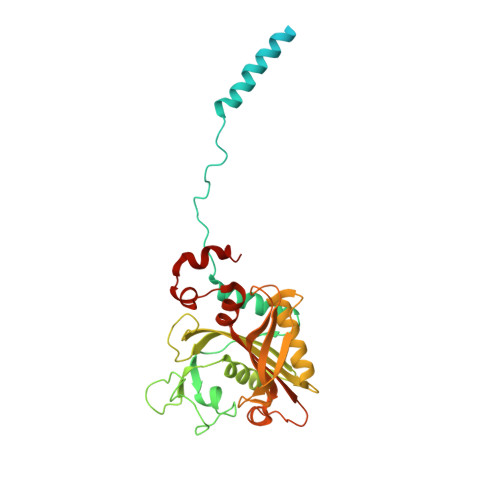
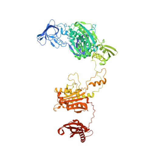
    7DAW, 7DB7, 7DB8

  • PubMed Abstract: 

    Mycobacteria tuberculosis (Mtb) remains the deadliest pathogenic bacteria worldwide. The search for new antibiotics to treat drug-sensitive as well as drug-resistant tuberculosis has become a priority. The essential enzyme phenylalanyl-tRNA synthetase (PheRS) is an antibacterial drug target because of the large differences between bacterial and human PheRS counterparts. In a high-throughput screening of 2148 bioactive compounds, PF-3845, which is a known inhibitor of human fatty acid amide hydrolase (FAAH), was identified inhibiting Mtb PheRS at K i ~0.73 ± 0.06 µM. The inhibition mechanism was studied with enzyme kinetics, protein structural modelling and crystallography, in comparison to a PheRS inhibitor of the noted phenyl-thiazolylurea-sulfonamide class. The 2.3-Å crystal structure of Mtb PheRS in complex with PF-3845 revealed its novel binding mode, in which a trifluoromethyl-pyridinylphenyl group occupies the Phe pocket while a piperidine-piperazine urea group binds into the ATP pocket through an interaction network enforced by a sulfate ion. It represents the first non-nucleoside bi-substrate competitive inhibitor of bacterial PheRS. PF-3845 inhibits the in vitro growth of Mtb H37Rv at ~24 µM, and the potency of PF-3845 increased against Mtb pheS-FDAS, suggesting on target activity in mycobacterial whole cells.  PF-3845 does not inhibit human cytoplasmic or mitochondrial PheRS in biochemical assay, which can be explained from the crystal structures. Further medicinal chemistry efforts focused on the piperidine-piperazine urea moiety may result in the identification of a selective antibacterial lead compound.


  • Organizational Affiliation
    • Global Health Drug Discovery Institute, China.

Macromolecule Content 

  • Total Structure Weight: 129.19 kDa 
  • Atom Count: 8,683 
  • Modeled Residue Count: 1,103 
  • Deposited Residue Count: 1,186 
  • Unique protein chains: 2

Macromolecules

Find similar proteins by:|  3D Structure
Entity ID: 1
MoleculeChains  Sequence LengthOrganismDetailsImage
Phenylalanine--tRNA ligase alpha subunit341Mycobacterium tuberculosis H37RvMutation(s): 0 
Gene Names: pheSRv1649MTCY06H11.14
EC: 6.1.1.20
UniProt
Find proteins for P9WFU3 (Mycobacterium tuberculosis (strain ATCC 25618 / H37Rv))
Explore P9WFU3 
Go to UniProtKB:  P9WFU3
Entity Groups
Sequence Clusters30% Identity50% Identity70% Identity90% Identity95% Identity100% Identity
UniProt GroupP9WFU3
Sequence Annotations
Expand
Reference Sequence
Find similar proteins by:|  3D Structure
Entity ID: 2
MoleculeChains  Sequence LengthOrganismDetailsImage
Phenylalanine--tRNA ligase beta subunit845Mycobacterium tuberculosis H37RvMutation(s): 0 
Gene Names: pheTRv1650MTCY06H11.15
EC: 6.1.1.20
UniProt
Find proteins for P9WFU1 (Mycobacterium tuberculosis (strain ATCC 25618 / H37Rv))
Explore P9WFU1 
Go to UniProtKB:  P9WFU1
Entity Groups
Sequence Clusters30% Identity50% Identity70% Identity90% Identity95% Identity100% Identity
UniProt GroupP9WFU1
Sequence Annotations
Expand
Reference Sequence

Small Molecules

Ligands 2 Unique
IDChains Name / Formula / InChI Key2D Diagram3D Interactions
H2L
(Subject of Investigation/LOI)

Query on H2L


Download:Ideal Coordinates CCD File
C [auth A]1-[3-[2-(1H-indol-3-yl)ethylsulfamoyl]phenyl]-3-(1,3-thiazol-2-yl)urea
C20 H19 N5 O3 S2
NYYVMDXWTXBRNW-UHFFFAOYSA-N
SO4

Query on SO4


Download:Ideal Coordinates CCD File
D [auth A]
E [auth A]
F [auth B]
G [auth B]
H [auth B]
D [auth A],
E [auth A],
F [auth B],
G [auth B],
H [auth B],
I [auth B],
J [auth B],
K [auth B],
L [auth B],
M [auth B],
N [auth B],
O [auth B]
SULFATE ION
O4 S
QAOWNCQODCNURD-UHFFFAOYSA-L

Experimental Data & Validation

Experimental Data

  • Method: X-RAY DIFFRACTION
  • Resolution: 2.71 Å
  • R-Value Free:  0.219 (Depositor), 0.218 (DCC) 
  • R-Value Work:  0.193 (Depositor), 0.194 (DCC) 
  • R-Value Observed: 0.194 (Depositor) 
Space Group: C 2 2 21
Unit Cell:
Length ( Å )Angle ( ˚ )
a = 268.38α = 90
b = 298.801β = 90
c = 65.712γ = 90
Software Package:
Software NamePurpose
PHENIXrefinement
PHENIXphasing
Cootmodel building
HKL-2000data reduction
HKL-2000data scaling

Structure Validation

View Full Validation Report


Ligand Structure Quality Assessment 


Entry History 

Deposition Data

Revision History  (Full details and data files)

  • Version 1.0: 2021-01-13
    Type: Initial release
  • Version 1.1: 2021-01-20
    Changes: Database references
  • Version 1.2: 2023-11-29
    Changes: Data collection, Database references, Refinement description