7D98 | pdb_00007d98

Crystal structure of full-length CbnR complexed with the target DNA complex


Experimental Data Snapshot

  • Method: X-RAY DIFFRACTION
  • Resolution: 3.60 Å
  • R-Value Free: 
    0.286 (Depositor), 0.287 (DCC) 
  • R-Value Work: 
    0.234 (Depositor), 0.237 (DCC) 
  • R-Value Observed: 
    0.237 (Depositor) 

Starting Model: experimental
View more details

wwPDB Validation 3D Report Full Report

Validation slider image for 7D98

This is version 1.2 of the entry. See complete history

Literature

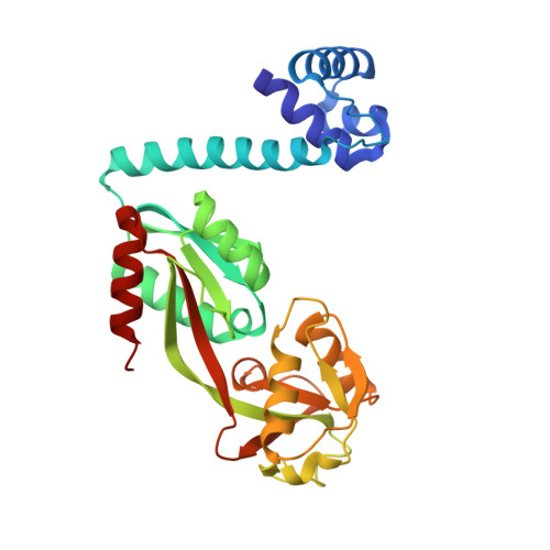
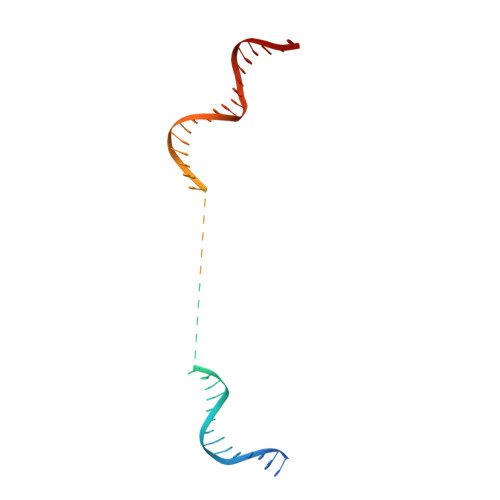
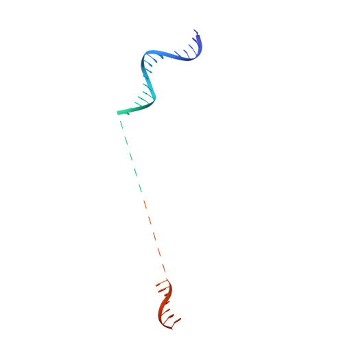
Crystal structure of the full-length LysR-type transcription regulator CbnR in complex with promoter DNA.

Giannopoulou, E.A.Senda, M.Koentjoro, M.P.Adachi, N.Ogawa, N.Senda, T.

(2021) FEBS J 288: 4560-4575

  • DOI: https://doi.org/10.1111/febs.15764
  • Primary Citation Related Structures: 
    7D98

  • PubMed Abstract: 

    LysR-type transcription regulators (LTTRs) comprise one of the largest families of transcriptional regulators in bacteria. They are typically homo-tetrameric proteins and interact with promoter DNA of ~ 50-60 bp. Earlier biochemical studies have suggested that LTTR binding to promoter DNA bends the DNA and, upon inducer binding, the bend angle of the DNA is reduced through a quaternary structure change of the tetrameric LTTR, leading to the activation of transcription. To date, crystal structures of full-length LTTRs, DNA-binding domains (DBD) with their target DNAs, and the regulatory domains with and without inducer molecules have been reported. However, these crystal structures have not provided direct evidence of the quaternary structure changes of LTTRs or of the molecular mechanism underlying these changes. Here, we report the first crystal structure of a full-length LTTR, CbnR, in complex with its promoter DNA. The crystal structure showed that, in the absence of bound inducer molecules, the four DBDs of the tetrameric CbnR interact with the promoter DNA, bending the DNA by ~ 70°. Structural comparison between the DNA-free and DNA-bound forms demonstrates that the quaternary structure change of the tetrameric CbnR required for promoter region-binding arises from relative orientation changes of the three domains in each subunit. The mechanism of the quaternary structure change caused by inducer binding is also discussed based on the present crystal structure, affinity analysis between CbnR and the promoter DNA, and earlier mutational studies on CbnR. DATABASE: Atomic coordinates and structure factors for the full-length Cupriavidus necator NH9 CbnR in complex with promoter DNA are available in the Protein Data Bank under the accession code 7D98.


  • Organizational Affiliation
    • Structural Biology Research Center, Photon Factory, Institute of Materials Structure Science, High Energy Accelerator Research Organization (KEK), Tsukuba, Japan.

Macromolecule Content 

  • Total Structure Weight: 162.85 kDa 
  • Atom Count: 9,258 
  • Modeled Residue Count: 1,201 
  • Deposited Residue Count: 1,288 
  • Unique protein chains: 1
  • Unique nucleic acid chains: 2

Macromolecules


Find similar proteins by:|  3D Structure
Entity ID: 1
MoleculeChains  Sequence LengthOrganismDetailsImage
LysR-type regulatory proteinA,
B,
C [auth P],
D [auth Q]
294Cupriavidus necatorMutation(s): 0 
Gene Names: cbnRBJN34_37375
UniProt
Find proteins for Q9WXC7 (Cupriavidus necator)
Explore Q9WXC7 
Go to UniProtKB:  Q9WXC7
Entity Groups
Sequence Clusters30% Identity50% Identity70% Identity90% Identity95% Identity100% Identity
UniProt GroupQ9WXC7
Sequence Annotations
Expand
Reference Sequence
Find similar nucleic acids by:  (by identity cutoff) 
Entity ID: 2
MoleculeChains LengthOrganismImage
DNA (56-mer)E [auth G]56Cupriavidus necator
Sequence Annotations
Expand
Reference Sequence
Find similar nucleic acids by:  (by identity cutoff) 
Entity ID: 3
MoleculeChains LengthOrganismImage
DNA (56-mer)F [auth H]56Cupriavidus necator
Sequence Annotations
Expand
Reference Sequence

Experimental Data & Validation

Experimental Data

  • Method: X-RAY DIFFRACTION
  • Resolution: 3.60 Å
  • R-Value Free:  0.286 (Depositor), 0.287 (DCC) 
  • R-Value Work:  0.234 (Depositor), 0.237 (DCC) 
  • R-Value Observed: 0.237 (Depositor) 
Space Group: P 61 2 2
Unit Cell:
Length ( Å )Angle ( ˚ )
a = 105.819α = 90
b = 105.819β = 90
c = 602.516γ = 120
Software Package:
Software NamePurpose
XDSdata processing
Aimlessdata scaling
Cootmodel building
MOLREPphasing
PHENIXrefinement

Structure Validation

View Full Validation Report


Entry History 

& Funding Information

Deposition Data


Funding OrganizationLocationGrant Number
Japan Society for the Promotion of Science (JSPS)Japan18K06098
Japan Agency for Medical Research and Development (AMED)JapanJP17am0101001

Revision History  (Full details and data files)

  • Version 1.0: 2021-03-10
    Type: Initial release
  • Version 1.1: 2021-08-18
    Changes: Database references
  • Version 1.2: 2023-11-29
    Changes: Data collection, Refinement description