


Crystal Structure of double mutant Y115E Y117E human Secretory Glutaminyl Cyclase in complex with LSB-09
Dileep, K.V., Ihara, K., Sakai, N., Shirozu, M., Zhang, K.Y.J.To be published.
Experimental Data Snapshot
Starting Model: experimental
View more details
Entity ID: 1 | |||||
|---|---|---|---|---|---|
| Molecule | Chains | Sequence Length | Organism | Details | Image |
| Glutaminyl-peptide cyclotransferase | 329 | Homo sapiens | Mutation(s): 2 Gene Names: QPCT EC: 2.3.2.5 | 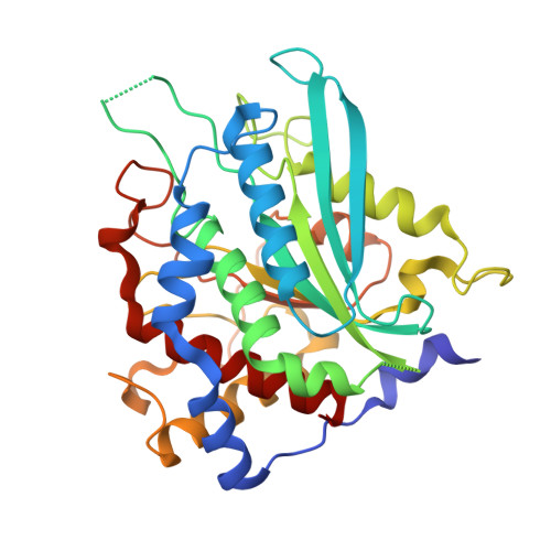 | |
UniProt & NIH Common Fund Data Resources | |||||
Find proteins for Q16769 (Homo sapiens) Explore Q16769 Go to UniProtKB: Q16769 | |||||
PHAROS: Q16769 GTEx: ENSG00000115828 | |||||
Entity Groups | |||||
| Sequence Clusters | 30% Identity50% Identity70% Identity90% Identity95% Identity100% Identity | ||||
| UniProt Group | Q16769 | ||||
Sequence AnnotationsExpand | |||||
| |||||
| Ligands 4 Unique | |||||
|---|---|---|---|---|---|
| ID | Chains | Name / Formula / InChI Key | 2D Diagram | 3D Interactions | |
| GYO (Subject of Investigation/LOI) Query on GYO | L [auth A], S [auth B], Y [auth C] | ~{N}-[(1~{S},2~{S})-2-(2-methoxyphenyl)cyclopropyl]-3~{H}-benzimidazole-5-carboxamide C18 H17 N3 O2 ZKYVGLRMGWYWNE-ZFWWWQNUSA-N |  | ||
| SO4 Query on SO4 | I [auth A] J [auth A] K [auth A] Q [auth B] R [auth B] | SULFATE ION O4 S QAOWNCQODCNURD-UHFFFAOYSA-L |  | ||
| ZN Query on ZN | D [auth A], M [auth B], T [auth C] | ZINC ION Zn PTFCDOFLOPIGGS-UHFFFAOYSA-N |  | ||
| EDO Query on EDO | E [auth A] F [auth A] G [auth A] H [auth A] N [auth B] | 1,2-ETHANEDIOL C2 H6 O2 LYCAIKOWRPUZTN-UHFFFAOYSA-N |  | ||
| Length ( Å ) | Angle ( ˚ ) |
|---|---|
| a = 86.181 | α = 90 |
| b = 149.303 | β = 97.64 |
| c = 94.539 | γ = 90 |
| Software Name | Purpose |
|---|---|
| PHENIX | refinement |
| XDS | data reduction |
| Aimless | data scaling |
| PDB_EXTRACT | data extraction |
| PHASER | phasing |
| Funding Organization | Location | Grant Number |
|---|---|---|
| Japan Society for the Promotion of Science (JSPS) | Japan | 16F16385 |


