Structure-guided development of Pb 2+ -binding DNA aptamers.
Liu, H., Gao, Y., Mathivanan, J., Shen, F., Chen, X., Li, Y., Shao, Z., Zhang, Y., Shao, Q., Sheng, J., Gan, J.(2022) Sci Rep 12: 460-460
- PubMed: 35013452 Search on PubMedSearch on PubMed Central
- DOI: https://doi.org/10.1038/s41598-021-04243-2
- Primary Citation Related Structures:
7D31, 7D32, 7D33 - PubMed Abstract:
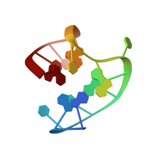
Owing to its great threat to human health and environment, Pb 2+ pollution has been recognized as a major public problem by the World Health Organization (WHO). Many DNA aptamers have been utilized in the development of Pb 2+ -detection sensors, but the underlying mechanisms remain elusive. Here, we report three Pb 2+ -complexed structures of the thrombin binding aptamer (TBA). These high-resolution crystal structures showed that TBA forms intramolecular G-quadruplex and Pb 2+ is bound by the two G-tetrads in the center. Compared to K + -stabilized G-quadruplexes, the coordinating distance between Pb 2+ and the G-tetrads are much shorter. The T3T4 and T12T13 linkers play important roles in dimerization and crystallization of TBA, but they are changeable for Pb 2+ -binding. In combination with mutagenesis and CD spectra, the G8C mutant structure unraveled that the T7G8T9 linker of TBA is also variable. In addition to expansion of the Pb 2+ -binding aptamer sequences, our study also set up one great example for quick and rational development of other aptamers with similar or optimized binding activity.
- Shanghai Public Health Clinical Center, State Key Laboratory of Genetic Engineering, Collaborative Innovation Center of Genetics and Development, School of Life Sciences, Fudan University, 2005 Songhu Road, Yangpu District, Shanghai, 200438, People's Republic of China.
Organizational Affiliation: