7CZB

The cryo-EM structure of the ERAD retrotranslocation channel formed by human Derlin-1


Experimental Data Snapshot

  • Method: ELECTRON MICROSCOPY
  • Resolution: 3.80 Å
  • Aggregation State: PARTICLE 
  • Reconstruction Method: SINGLE PARTICLE 

wwPDB Validation   3D Report Full Report


This is version 1.1 of the entry. See complete history


Literature

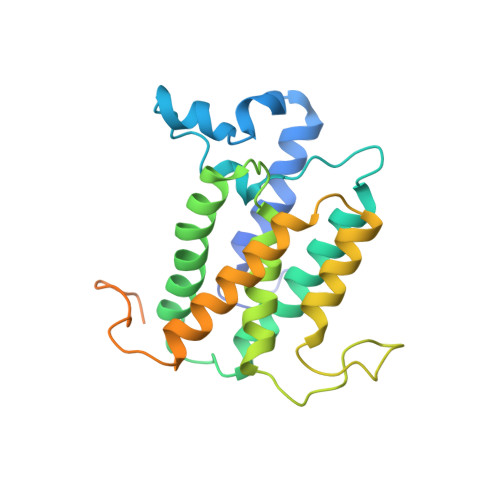
The cryo-EM structure of an ERAD protein channel formed by tetrameric human Derlin-1.

Rao, B.Li, S.Yao, D.Wang, Q.Xia, Y.Jia, Y.Shen, Y.Cao, Y.

(2021) Sci Adv 7

  • DOI: https://doi.org/10.1126/sciadv.abe8591
  • Primary Citation of Related Structures:  
    7CZB

  • PubMed Abstract: 

    Endoplasmic reticulum-associated degradation (ERAD) is a process directing misfolded proteins from the ER lumen and membrane to the degradation machinery in the cytosol. A key step in ERAD is the translocation of ER proteins to the cytosol. Derlins are essential for protein translocation in ERAD, but the mechanism remains unclear. Here, we solved the structure of human Derlin-1 by cryo-electron microscopy. The structure shows that Derlin-1 forms a homotetramer that encircles a large tunnel traversing the ER membrane. The tunnel has a diameter of about 12 to 15 angstroms, large enough to allow an α helix to pass through. The structure also shows a lateral gate within the membrane, providing access of transmembrane proteins to the tunnel, and thus, human Derlin-1 forms a protein channel for translocation of misfolded proteins. Our structure is different from the monomeric yeast Derlin structure previously reported, which forms a semichannel with another protein.


  • Organizational Affiliation

    CAS Center for Excellence in Molecular Cell Science, Shanghai Institute of Biochemistry and Cell Biology, Chinese Academy of Sciences, University of Chinese Academy of Sciences, 333 Haike Road, Shanghai 201210, China.


Macromolecules
Find similar proteins by:  (by identity cutoff)  |  3D Structure
Entity ID: 1
MoleculeChains Sequence LengthOrganismDetailsImage
Derlin-1
A, B, C, D
251Homo sapiensMutation(s): 0 
Gene Names: DERL1DER1UNQ243/PRO276
Membrane Entity: Yes 
UniProt & NIH Common Fund Data Resources
Find proteins for Q9BUN8 (Homo sapiens)
Explore Q9BUN8 
Go to UniProtKB:  Q9BUN8
PHAROS:  Q9BUN8
GTEx:  ENSG00000136986 
Entity Groups  
Sequence Clusters30% Identity50% Identity70% Identity90% Identity95% Identity100% Identity
UniProt GroupQ9BUN8
Sequence Annotations
Expand
  • Reference Sequence
Experimental Data & Validation

Experimental Data

  • Method: ELECTRON MICROSCOPY
  • Resolution: 3.80 Å
  • Aggregation State: PARTICLE 
  • Reconstruction Method: SINGLE PARTICLE 

Structure Validation

View Full Validation Report


Entry History & Funding Information

Deposition Data


Funding OrganizationLocationGrant Number
National Natural Science Foundation of China (NSFC)ChinaU1632132
National Natural Science Foundation of China (NSFC)China31670849
Ministry of Science and Technology (MoST, China)China2017YFC1001303
Ministry of Science and Technology (MoST, China)China2018YFC1004704

Revision History  (Full details and data files)

  • Version 1.0: 2021-03-17
    Type: Initial release
  • Version 1.1: 2024-03-27
    Changes: Data collection, Database references