


Crystal structure of Arabinose isomerase from hyper thermophilic hybrid AI8 with Adonitol
Hoang, N.K.Q., Dhanasingh, I., Cao, T.P., Sung, J.Y., Shin, S.M., Lee, D.W., Lee, S.H.To be published.
Experimental Data Snapshot
Starting Model: experimental
View more details
Entity ID: 1 | |||||
|---|---|---|---|---|---|
| Molecule | Chains | Sequence Length | Organism | Details | Image |
| L-arabinose isomerase | 497 | Geobacillus kaustophilus HTA426, Alicyclobacillus sp. TP7, Alicyclobacillus acidocaldarius subsp. acidocaldarius | Mutation(s): 0 Gene Names: araA, GK1904 EC: 5.3.1.4 | 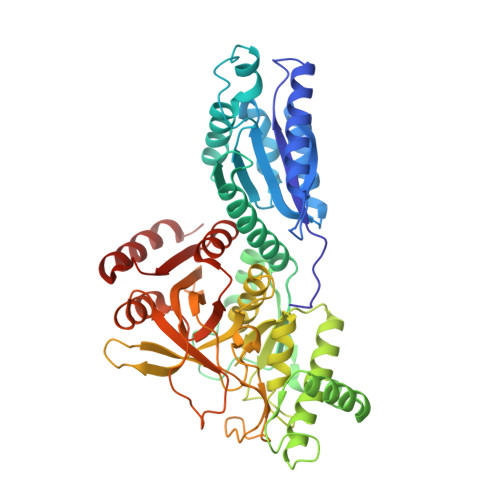 | |
UniProt | |||||
Entity Groups | |||||
| Sequence Clusters | 30% Identity50% Identity70% Identity90% Identity95% Identity100% Identity | ||||
| UniProt Groups | Q5KYP7K0IGW6Q2VMT2 | ||||
Sequence AnnotationsExpand | |||||
Reference Sequence | |||||
| Ligands 3 Unique | |||||
|---|---|---|---|---|---|
| ID | Chains | Name / Formula / InChI Key | 2D Diagram | 3D Interactions | |
| RB0 (Subject of Investigation/LOI) Download:Ideal Coordinates CCD File | J [auth B] O [auth D] P [auth D] S [auth E] U [auth F] | D-ribitol C5 H12 O5 HEBKCHPVOIAQTA-ZXFHETKHSA-N |  | ||
| MPD Download:Ideal Coordinates CCD File | G [auth A], M [auth C] | (4S)-2-METHYL-2,4-PENTANEDIOL C6 H14 O2 SVTBMSDMJJWYQN-YFKPBYRVSA-N |  | ||
| MN Download:Ideal Coordinates CCD File | H [auth A] I [auth A] K [auth B] L [auth B] N [auth C] | MANGANESE (II) ION Mn WAEMQWOKJMHJLA-UHFFFAOYSA-N |  | ||
| Length ( Å ) | Angle ( ˚ ) |
|---|---|
| a = 205.582 | α = 90 |
| b = 82.12 | β = 117.93 |
| c = 192.726 | γ = 90 |
| Software Name | Purpose |
|---|---|
| PHENIX | refinement |
| HKL-2000 | data reduction |
| HKL-2000 | data scaling |
| PDB_EXTRACT | data extraction |
| MOLREP | phasing |


