7CTO

Staphylococcus aureus MsrB


Experimental Data Snapshot

  • Method: X-RAY DIFFRACTION
  • Resolution: 3.47 Å
  • R-Value Free: 0.323 
  • R-Value Work: 0.216 
  • R-Value Observed: 0.222 

wwPDB Validation   3D Report Full Report


This is version 1.2 of the entry. See complete history


Literature

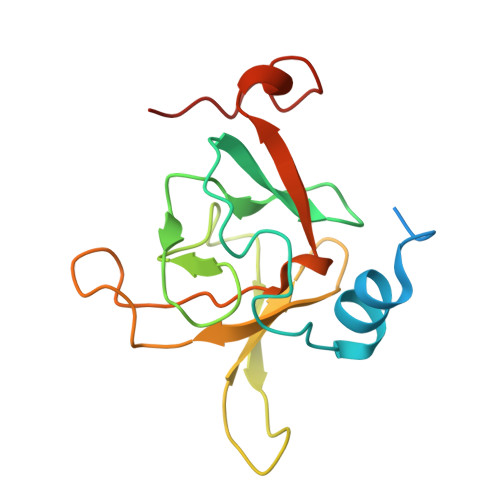
Implication of Staphylococcus aureus MsrB dimerization upon oxidation.

Kim, H.J.

(2020) Biochem Biophys Res Commun 533: 118-124

  • DOI: https://doi.org/10.1016/j.bbrc.2020.08.070
  • Primary Citation of Related Structures:  
    7CTO

  • PubMed Abstract: 

    Oxidative modification of protein structure has been shown to play a significant role in bacterial virulence and metabolism. The sulfur-containing residues are susceptible to oxidation and the enzymatic reversal of oxidized cysteine or methionine is detected in many organisms. Methionine sulfoxide reductases (Msr) are responsible for reducing oxidized methionine. The two different Msrs, MsrA and MsrB, reduce methionine R-sulfoxide and methionine S-sulfoxide, respectively through self-oxidation. This study elucidated the structure of MsrB from Staphylococcus aureus Mu50 and its changes upon oxidation. The active site shows two reduced cysteines in a close contact, implying disulfide bond would form without major structural rearrangement. When the protein is exposed to an oxidative condition, a dimeric state is observed. The dimerization of SA MsrB creates a valley structure for accepting peptidyl substrates. To the best of our knowledge, oxidation induced dimerization of SA MsrB would help to understand mechanism behind redox control that has not been well characterized.


  • Organizational Affiliation

    College of Pharmacy, Woosuk University, Wanju, 55338, Republic of Korea. Electronic address: hyojungkim@woosuk.ac.kr.


Macromolecules
Find similar proteins by:  (by identity cutoff)  |  3D Structure
Entity ID: 1
MoleculeChains Sequence LengthOrganismDetailsImage
Peptide methionine sulfoxide reductase MsrB
A, B, C, D, E
A, B, C, D, E, F
159Staphylococcus aureusMutation(s): 0 
Gene Names: 
EC: 1.8.4.12
UniProt
Find proteins for W8TTH3 (Staphylococcus aureus)
Explore W8TTH3 
Go to UniProtKB:  W8TTH3
Entity Groups  
Sequence Clusters30% Identity50% Identity70% Identity90% Identity95% Identity100% Identity
UniProt GroupW8TTH3
Sequence Annotations
Expand
  • Reference Sequence
Experimental Data & Validation

Experimental Data

  • Method: X-RAY DIFFRACTION
  • Resolution: 3.47 Å
  • R-Value Free: 0.323 
  • R-Value Work: 0.216 
  • R-Value Observed: 0.222 
  • Space Group: P 21 21 2
Unit Cell:
Length ( Å )Angle ( ˚ )
a = 114.825α = 90
b = 122.19β = 90
c = 73.27γ = 90
Software Package:
Software NamePurpose
REFMACrefinement
PDB_EXTRACTdata extraction
Aimlessdata reduction
Aimlessdata scaling
PHASERphasing

Structure Validation

View Full Validation Report


Entry History 

Deposition Data

  • Released Date: 2020-10-07 
  • Deposition Author(s): Kim, H.J.

Revision History  (Full details and data files)

  • Version 1.0: 2020-10-07
    Type: Initial release
  • Version 1.1: 2020-10-28
    Changes: Database references
  • Version 1.2: 2023-11-29
    Changes: Data collection, Database references, Refinement description