Structural basis of catalysis and substrate recognition by the NAD(H)-dependent alpha-d-glucuronidase from the glycoside hydrolase family 4.
Mohapatra, S.B., Manoj, N.(2021) Biochem J 478: 943-959
- PubMed: 33565573 Search on PubMed
- DOI: https://doi.org/10.1042/BCJ20200824
- Primary Citation Related Structures:
7CTD, 7CTL, 7CTM - PubMed Abstract:
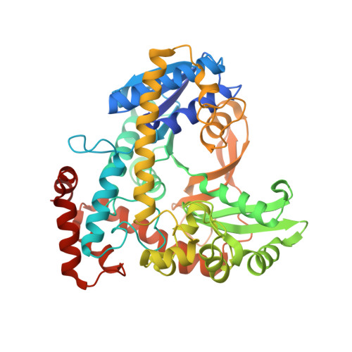
Members of the glycoside hydrolase family 4 (GH4) employ an unusual glycosidic bond cleavage mechanism utilizing NAD(H) and a divalent metal ion, under reducing conditions. These enzymes act upon a diverse range of glycosides, and unlike most other GH families, homologs here are known to accommodate both α- and β-anomeric specificities within the same active site. Here, we report the catalytic properties and the crystal structures of TmAgu4B, an α-d-glucuronidase from the hyperthermophile Thermotoga maritima. The structures in three different states include the apo form, the NADH bound holo form, and the ternary complex with NADH and the reaction product d-glucuronic acid, at 2.15, 1.97 and 1.85 Å resolutions, respectively. These structures reveal the step-wise route of conformational changes required in the active site to achieve the catalytically competent state, and illustrate the direct role of residues that determine the reaction mechanism. Furthermore, a structural transition of a helical region in the active site to a turn geometry resulting in the rearrangement of a unique arginine residue governs the exclusive glucopyranosiduronic acid recognition in TmAgu4B. Mutational studies show that modifications of the glycone binding site geometry lead to catalytic failure and indicate overlapping roles of specific residues in catalysis and substrate recognition. The data highlight hitherto unreported molecular features and associated active site dynamics that determine the structure-function relationships within the unique GH4 family.
- Department of Biotechnology, Bhupat and Jyoti Mehta School of Biosciences, Indian Institute of Technology Madras, Chennai 600036, India.
Organizational Affiliation: